
Volume 26 Issue 6, June 2021
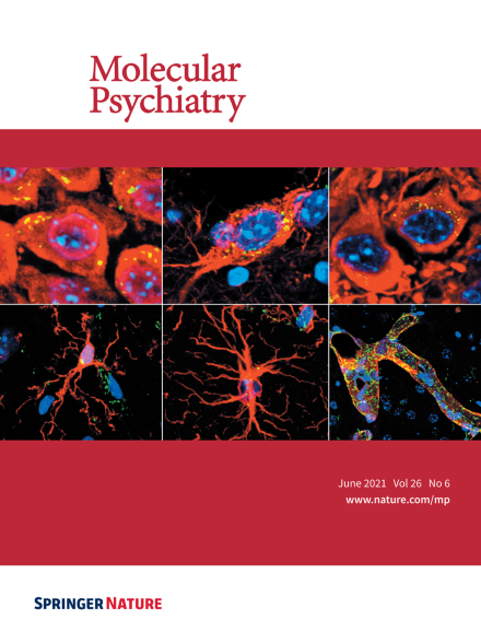
Immunohistochemical localization of TSPO protein in neuronal and non-neuronal hippocampal cells of adult mice using immunofluorescence staining. The photomicrographs show representative Z-stack images acquired through confocal microscopy, with nuclear staining (DAPI) in blue, TSPO in green and various CNS cells of interest in red. TSPO co-localizing with the cellular markers of interest appears in yellow. Top row images show TSPO co-localizing with the post-mitotic neuronal markers NeuN (left image), SMI-32 (middle image) and MAP-2 (right image). Lower row images show non-neuronal TSPO protein expression, including expression in Iba1-positive microglia (left image), GFAP-positive astrocytes (middle image) and Glut1-positive vascular endothelial cells (right image).For more information see the article by Tina Notter et al. on page 2025-2037.
Image
-
Advertisement
