
Volume 26 Issue 5, May 2021
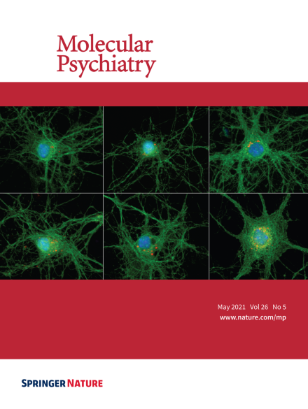
Immunofluorescent staining of neuron-microglia co-cultures. Images show neurons from Pten WT/WT and from Pten autism mouse model Pten WT/m3m4 , and Pten m3m4/m3m (Left to right) co-cultured with Pten WT/WT microglia (Top panels) and Pten m3m4/m3m microglia (Bottom panels). Neurons are labeled green with Tuj1; C1q (red) punctae show accumulation on neuronal surfaces. For more information see the article by Prof. Charis Eng et al. on page 1458-1471.
Image
-
Advertisement
