Thank you for visiting nature.com. You are using a browser version with limited support for CSS. To obtain
the best experience, we recommend you use a more up to date browser (or turn off compatibility mode in
Internet Explorer). In the meantime, to ensure continued support, we are displaying the site without styles
and JavaScript.
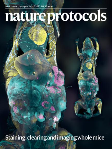
Nanobody(VHH)-boosted 3D imaging of solvent-cleared organs (vDISCO) enables the labelling of single cells in intact animals or whole organs. The cover shows a vDISCO image of a whole mouse where individual metastatic cells (magenta), bones and organs (cyan) and muscles (yellow) are labelled via nanobodies.
In this Perspective, authors from the UK Consortium on Metabolic Phenotyping propose a ‘fit-for-purpose’, four-tiered framework to evaluate the reliability of targeted metabolomics analyses, addressing the need for community-accepted, harmonized guidelines for tiers other than full validation.
This Protocol Extension presents an updated tetrahedral DNA nanostructure for the delivery of various bioactive molecules that enables an active targeting strategy to be used for stimuli-sensitive conformation changes and on-site cargo release.
The multi-attribute method directly characterizes and monitors multiple product quality attributes of a biopharmaceutical product via proteolytic digestion of the sample followed by reversed-phase chromatographic separation and high-resolution liquid chromatography-mass spectrometry analysis.
Single-cell screening and sorting is useful for addressing many biological questions. The high-throughput droplet analyzer and sorter described in this protocol has been designed so that it can be constructed without specialist engineering training.
A surgical procedure that preserves hearing while creating an imaging window in the mouse cochlea, enabling the functional imaging of cochlear cells in vivo.
Mesoporous carbonaceous nanomaterials have technological potential for diverse applications ranging from gas separation to energy storage. We describe the template-directed assembly of monomicelles to make mesoporous materials with different structures.
The amplification of misfolded alpha-synuclein aggregates in vitro can be used for the detection, via fluorescent dyes, of pathologic amyloids in cerebrospinal fluid samples from patients affected by Parkinson’s disease, dementia with Lewy bodies or multiple-system atrophy.
A full-body nanobody-based immunolabeling and clearing method that renders mice transparent in 3 weeks, enhancing the signal of fluorescent proteins and allowing their reliable detection and quantification, at cellular resolution, within an entire body.
Making microfluidic chips using 3D printers allows rapid fabrication of ready-to-use products from digital 3D designs with minimal human intervention. This ‘print–pause–print’ protocol describes how to make transparent, multimaterial chips.
A protocol for quantitative genome-wide detection of replication initiation, fork progression and termination, based on purification and strand-specific sequencing of Okazaki fragments isolated from mammalian or yeast cells.
The tandem mass spectrometers used in clinical chemistry are expensive. This protocol describes how to generate similar results using a single mass spectrometry detector by optimizing in-source fragmentation and data analysis via correlated ion monitoring.
We present a protocol for the synthesis and usage of QM-FN-SO3, a near-infrared aggregation-induced-emission-active fluorescent probe capable of crossing the blood–brain barrier and ultrasensitively lighting up amyloid-β plaques in living mice.
The authors present a protocol for the modular 3D bioengineering of multilineage skeletal muscles from human induced pluripotent stem cells, along with assays to characterize morphological and functional features of the artificial muscle constructs.