
Volume 18 Issue 5, May 2023
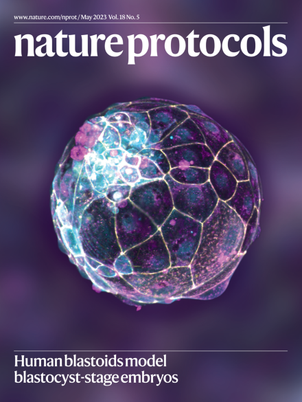
Human blastoids model blastocyst-stage embryos
The cover shows a confocal image of a human blastoid labeled for the tight junction molecule ZO-1 (yellow), the adherens junction molecule CDH1 (magenta), the apical domain molecule aPKC (cyan) and nuclei (blue).
Image: Alok Javali, Nicolas Rivron group, Institute of Molecular Biotechnology of the Austrian Academy of Sciences (IMBA). Cover design: S. Whitham
Protocol Extensions
-
Advertisement
