
Volume 18 Issue 12, December 2023
An adaptive optics two-photon microscopy image of layer 4 of the primary somatosensory cortex
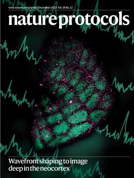
A composite image showing the barrel structure of the primary somatosensory cortex in mice delineated by thalamocortical boutons virally labeled with iGluSnFR3-v857.GPI (green) via injection into the ventral posteromedial thalamus. The vasculature is labeled via Cy5.5 in the bloodstream (magenta). The traces depict single-trial recordings of glutamate release at individual synapses in response to a 5-Hz train of air puffs to the vibrissae. See Yao et al.
Image: Rui Liu, University of California, San Diego Cover design: S. Whitham
Perspectives
-
Advertisement
