
Thank you for visiting nature.com. You are using a browser version with limited support for CSS. To obtain the best experience, we recommend you use a more up to date browser (or turn off compatibility mode in Internet Explorer). In the meantime, to ensure continued support, we are displaying the site without styles and JavaScript.

Volume 28 Issue 9, September 2023

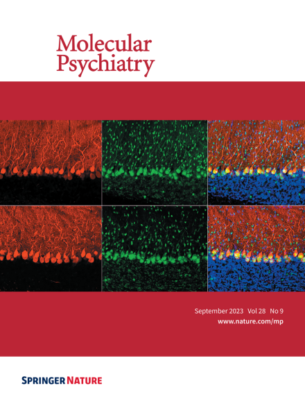
Confocal microscopy images of mouse cerebellar lobule V sections immunolabeled for calbindin D-28k (red), Fox2(green), and counterstained with DAPI (blue). The top row is from a wild-type mouse and the bottom row from a Bmal1-/- mouse. For more information see the article by Liu et al. on pages 3727–3738.

Image

Top of page ⤴

Correspondence

Top of page ⤴

Perspective

Top of page ⤴

Review Article

Top of page ⤴

Expert Review

Top of page ⤴

Systematic Review

Top of page ⤴

Article

Top of page ⤴

Search

Quick links