Thank you for visiting nature.com. You are using a browser version with limited support for CSS. To obtain
the best experience, we recommend you use a more up to date browser (or turn off compatibility mode in
Internet Explorer). In the meantime, to ensure continued support, we are displaying the site without styles
and JavaScript.
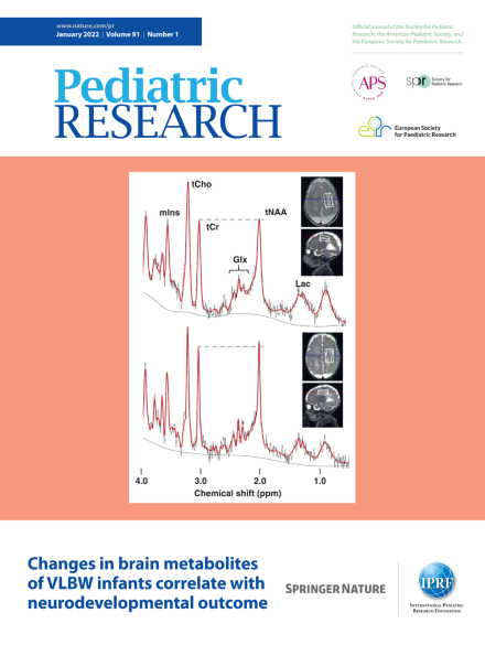
Magnetic resonance spectroscopy at term of very-low-birthweight infants with delayed (top) and normal (bottom) development. See article by Tomiyasu et al. on page 197.
A continuous oxygen supply is vital for fueling fetal growth, yet we know surprisingly little as to the precise mechanisms by which hypoxia reduces fetal growth. Animal models, while important, are limited by the enormous variability in mammalian physiologic responses to pregnancy. One of the earliest models for understanding hypoxia-induced reductions in fetal growth came from studies at high altitude that showed high altitude to have one of the strongest depressant effects on fetal growth. But all populations are not equally affected. Dolma et al. have added valuable information now showing that that Ladakhis, like Tibetans and Andeans, have a lower-than-expected frequency of small-for-gestational age (SGA) infants. Consistent with previous reports, these authors also found that uterine artery diameters at mid pregnancy (weeks 26–28) were larger in women giving birth to appropriate-for-gestational age (AGA) than SGA infants at high but, interestingly, not at low altitude. Much remains to be learned about the physiologic pathways by which hypoxia impairs fetal growth. The variability among high-altitude populations and increasing sophistication of tools for investigating causal mechanisms have the potential to expand our presently limited means for identifying new treatments for hypoxia-related complications of pregnancy and fetal life.