Volume 17 Issue 10, October 2022
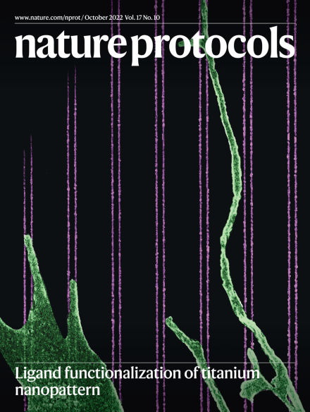
Fibroblast cell forming filopodia on nanopatterns
Scanning electron microscope image (pseudo-color) of a fibroblast cell forming filopodia along nanofibers functionalized by the integrin-binding peptide Arg–Gly–Asp (RGD). The nanopatterns are arrays of 20-nm-wide lines with 80-nm center-to-center distance in each line pair. Functionalization of ligands on titanium nanopatterns enables super-resolution fluorescence microscopy to study cell–ligand interactions at the molecular scale.
See Jain et al.
Image: Haogang Cai, New York University Grossman School of Medicine. Cover design: S. Harris