Volume 17
-
No. 12 December 2022
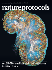
Human kidney development visualized in eight colors.3D rendering of an eight-color confocal image of a human fetal kidney labeled for nephrogenic and structural markers using the multispectral large-scale single-cell resolution 3D (mLSR-3D) imaging protocol by van Ineveld, Collot and colleagues featured in this issue of Nature Protocols.
See van Ineveld et al.
-
No. 11 November 2022
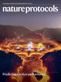
Constructing and interpreting molecular volcano plotsVolcano plots are a unique tool that reveal the optimal homogeneous catalyst(s) for a chemical reaction, which appear at (or near) the volcano summit or plateau. In this protocol, we provide a detailed overview of how volcanoes can be constructed and interpreted from the results of electronic structure computations. A standalone code, volcanic, that automates construction is also presented.
See Laplaza et al.
-
No. 10 October 2022
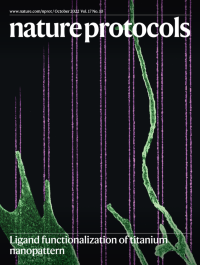
Fibroblast cell forming filopodia on nanopatternsScanning electron microscope image (pseudo-color) of a fibroblast cell forming filopodia along nanofibers functionalized by the integrin-binding peptide Arg–Gly–Asp (RGD). The nanopatterns are arrays of 20-nm-wide lines with 80-nm center-to-center distance in each line pair. Functionalization of ligands on titanium nanopatterns enables super-resolution fluorescence microscopy to study cell–ligand interactions at the molecular scale.
See Jain et al.
-
No. 9 September 2022
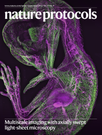
Ten-day-old chicken embryo imaged with a mesoSPIM light-sheet microscopeA ten-day-old chicken embryo cleared using benzyl benzoate and imaged using a mesoSPIM light-sheet microscope with axially swept excitation. Neurofilament is shown in green and autofluorescence in magenta.
See Dean et al.
-
No. 8 August 2022
Mnemiopsis leidyiAdult Mnemiopsis leidyi in culture.
See Presnell et al.
-
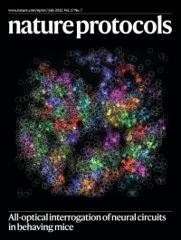
No. 7 July 2022
Targeted photostimulation of functionally defined ensembles of neuronsThe cover shows a strategy for ‘all-optical’ interrogation of neural circuits in vivo. Neuronal responses measured using two-photon calcium imaging were elicited by sequential, targeted two-photon photostimulation of 76 spatially clustered groups of neurons in mouse visual cortex in vivo. This large-scale but targeted activation is used to map the functional responses of neurons in the field of view for subsequent experiments. Responses are color-coded by the neurons targeted in a single group.
See Russell et al.
-
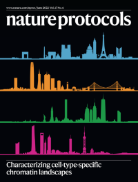
No. 6 June 2022
Characterizing cell-type-specific chromatin landscapesATAC-seq profiles the gene regulatory ‘landscape’ of a cell. Promoters, enhancers and other putative gene regulatory elements are identified as peaks in the data and these signals are highly cell-type-specific. Here, the skylines of Paris, San Francisco, New York City and Shanghai are depicted in the style of cell-type-specific ATAC-seq peaks.
See Grandi et al.
-
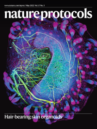
No. 5 May 2022
Hair-bearing skin organoidsThe image displays a human-stem-cell-derived skin organoid with radially growing hair follicles (green) and nerves (red/yellow). This particular organoid was grown from a genetically modified stem cell line where the Desmoplakin gene/protein is tagged with a GFP. As a consequence, the numerous desmosomes (cell–cell junctions) in the epidermis of the organoid glow green. Neurons are labeled with an antibody for beta-III tubulin.
See Lee et al.
-
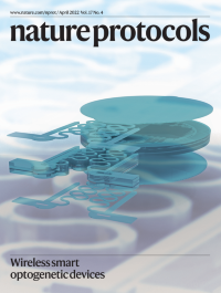
No. 4 April 2022
Wireless active optogenetic device with real-time stimulation controlThe layered schematics of a wireless optogenetic device capable of full subdermal implantation over the skull with real-time control over optical modulation on neural activities. Application of such devices in live rodent models enables profound behavior studies that explore the underlying neural principles behind outcome behaviors.
See Yang et al.
-
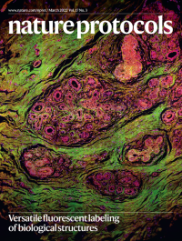
No. 3 March 2022
Human breast tissue stained with FLAREFormalin-fixed paraffin-embedded human breast tissue that was rapidly stained by FLARE (fluorescent labeling of abundant reactive entities), enabling informative visualization analogous to classic histology stains and holding the potential to revolutionize the clinical practice of examining tissue samples.
See Lee et al.
-
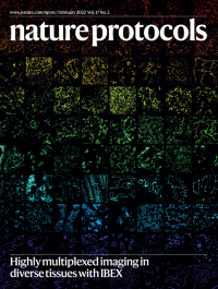
No. 2 February 2022
Multiplexed imaging of diverse human tissuesRainbow collage of IBEX images from nine different human tissues, including the lymph node, thymus, spleen, jejunum, kidney, liver, skin and heart. Individual images display unique cell types and anatomical structures defined by a single protein biomarker. All protein biomarkers are targeted by commercially available antibodies and obtained by IBEX, an open-source multiplexed imaging method.
See Radtke et al.
-
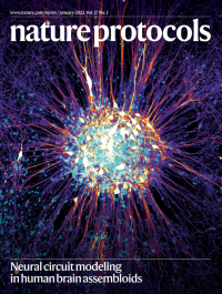
No. 1 January 2022
Human neurons in cultureImage of human striatal organoids that are derived from pluripotent stem cells in vitro and can be integrated with cortical cells to form cortico-striatal assembloids.
See Miura et al.