Thank you for visiting nature.com. You are using a browser version with limited support for CSS. To obtain
the best experience, we recommend you use a more up to date browser (or turn off compatibility mode in
Internet Explorer). In the meantime, to ensure continued support, we are displaying the site without styles
and JavaScript.
Imprinting effector and stem-like memory cell fates
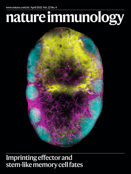
Groom and colleagues have developed a method to quantify a cell’s 3D location to determine the spatial positioning that directs T cell fate. Lightsheet imaging revealed that intranodal migration into distinct niches determines CD8+ T cell differentiation following viral infection.
Taiwan has leveraged its experience in containing the 2003 SARS outbreak by responding to the COVID-19 crisis with rapid measures, early deployment, prudent actions and transparency. Collectively, these actions have created the ‘Taiwan model’ for combating COVID-19.
HIV-1 infection of CD4+ T cells triggers the interaction of the mitochondrial proteins NLRX1 and FASTKD5 to promote oxidative phosphorylation, leading to increased viral replication. It has now been shown that this process can be blocked by metformin.
In the colonic environment, sustained Wnt–β-catenin activation in regulatory T cells promotes epigenetic rewiring toward proinflammatory RORγt+ Treg cells, whose expansion parallels the disease progression from inflammatory bowel disease (IBD) to manifest colorectal cancer (CRC).
The eradication of pathogens and establishment of immunological memory depend on the generation of both effector and long-lived memory cells within specialized immune niches. Whole-organ imaging demonstrates that, during viral infection, the fate of CD8+ T cells in lymph nodes is coupled to chemotactic signals controlling their distribution within different microanatomical sites.
In the midst of resurging COVID-19 cases, the second NIH/FDA virtual COVID-19 and Cytokines symposium was held on 1 December 2020, focusing on longitudinal studies of COVID-19 immunity, including long-term consequences, potential associations with autoimmunity and the multisystem inflammatory syndrome in children (MIS-C).
Ting and colleagues use multi-omics to examine the alterations undergone by CD4+ T cells following HIV-1 infection. They describe mechanistic changes that lead to elevated oxidative phosphorylation, which, if inhibited, leads to suppression of HIV-1 infection.
T cells are highly dynamic and their spatial and cellular interactions can influence their differentiation program. Groom and colleagues use three-dimensional spatial imaging to show that effector and stem-like memory cell fates are imposed within distinct lymph node regions.
Mucida and colleagues examine how the gut epithelial microenvironment alters CD4+ T cells during their conversion into intraepithelial lymphocytes. They reveal a stepwise process involving chromatin accessibility and transcription changes triggered by ThPOK downregulation.
The E3 ubiquitin ligase MDM2 inhibits the tumor suppressor p53 and is an important therapeutic target. Zou and colleagues demonstrate that MDM2 also has a T cell-intrinsic role that supports antitumor responses.
Gounari and colleagues examine how the Wnt–β-catenin signaling axis in regulatory T cells promotes inflammatory bowel disease and colonic dysplasia. Activated β-catenin induces epigenetic changes that alter expression of genes co-regulated by Foxp3 and TCF-1.
Sharma and colleagues identify the kinase DAPK3 as a positive regulator of the STING–interferon-β activation pathway. DAPK3 acts to modify E3 ubiquitin ligases that regulate STING K63-linked poly-ubiquitination.
Viral pathogens frequently target host cell antigen-processing pathways, including MHC-I–TAP peptide transporters, to evade host immunity. Blander and colleagues describe how MHC-I molecules can still cross-present antigen by re-routing ERGIC-resident MHC molecules to phagosomal vesicles, where phagolysosomal proteases act to shape the peptide repertoire for MHC-I presentation.
Kollias and colleagues examine the development of fibroblastic reticular cells (FRCs) during lymphoid organogenesis in gut Peyer’s patches (PPs). They show that PP FRCs develop from two separate mesenchymal cell lineages, which converge and form mosaic microenvironments that support immune cell activation and maintain intestinal homeostasis.
Growing evidence suggests that immune dysregulation is involved in the pathogenesis of myelodysplastic syndromes (MDSs). Glimcher and colleagues report haplosufficiency of the serine–threonine kinase RIOK2 leads to increased IL-22 production that, in turn, suppresses erythropoiesis. Blocking IL-22 rescues this defect in mice, suggesting that IL-22 blockade might be of therapeutic value in treating MDSs.