
Volume 32 Issue 2, February 2019
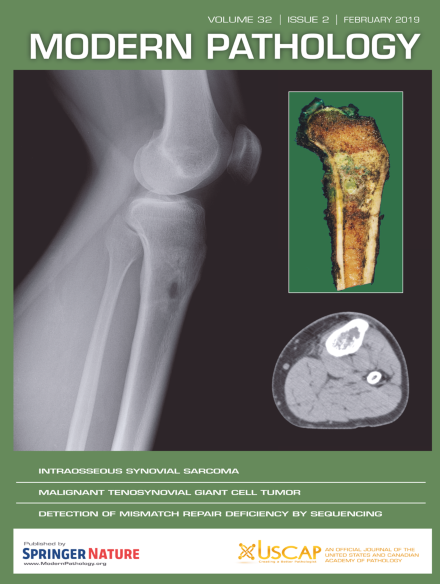
In this issue, The cover shows pathologic and radiographic findings of intraosseous synovial sarcoma. There is a fleshy intramedullary lesion in the metaphysis without a significant soft tissue component. Conventional lateral radiograph and axial CT showed a subtle mixed lytic-sclerotic lesion in the tibial metaphysis; the circumscribed oval lytic lesion on the conventional radiograph is a biopsy cavity. For more information, see the paper by Horvai et al, p231, this issue.
Inside the USCAP Journals
-
Advertisement
