
Volume 91 Issue 5, May 2011
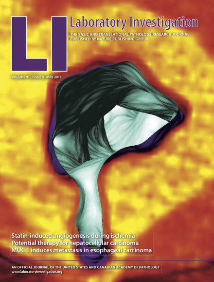
The cover shows a transverse sectional plane at branch point of reconstructed high resolution CT scan through abdominal aortic wall (denser yellow) from dog with mucopolysaccharidosis-I, looking down the aortic lumen. Intimal plaques associated with branch points bulge inward and taper distally. For more information see the paper by Lyons et al on page 665.
Inside Lab Invest
-
Advertisement
