
Thank you for visiting nature.com. You are using a browser version with limited support for CSS. To obtain the best experience, we recommend you use a more up to date browser (or turn off compatibility mode in Internet Explorer). In the meantime, to ensure continued support, we are displaying the site without styles and JavaScript.

Volume 14 Issue 5, May 2018

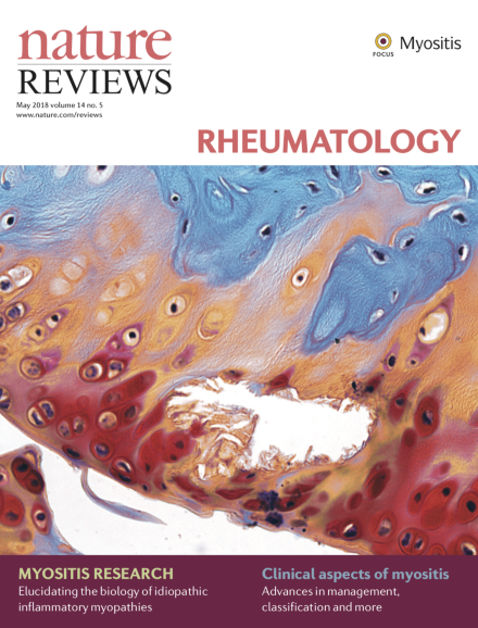
Image supplied by Dr Michal Dudek from the Faculty of Life Sciences, University of Manchester, Manchester, UK. The image shows knee articular cartilage from a chondrocyte-specific Bmal1-knockout mouse. The tissue was stained with safranin O and fast green. Deletion of the transcription factor brain and muscle Arnt-like protein 1 (BMAL1, also known as aryl hydrocarbon receptor nuclear translocator-like protein 1), a core component of the circadian clock, results in the loss of circadian rhythm and leads to degeneration of knee cartilage. The circadian clock controls the rhythmic expression of several hundred genes in cartilage and its function can be affected by inflammation and ageing, both of which are risk factors for osteoarthritis. Studies of the circadian clock will help us better understand cartilage physiology in health and disease.

Research Highlight

Top of page ⤴

In Brief

Top of page ⤴

Research Highlight

Top of page ⤴

News & Views

  • What causes muscle fibre necrosis in necrotizing myopathy? The authors of a new study propose that specific autoantibodies contribute to disease via immune-mediated processes; but given that these processes also occur in muscular dystrophies, how likely is that these antibodies trigger muscle fibre necrosis?

    • Marinos C. Dalakas
    News & Views
  • Interstitial lung disease (ILD) is a major cause of death and disability in patients with polymyositis or dermatomyositis. To begin to improve patient outcomes, the clinical, demographic and therapeutic factors associated with poor prognosis for patients with ILD associated with polymyositis or dermatomyositis need to be identified.

    • Sonye K. Danoff
    News & Views
Top of page ⤴

Review Article

  • Multiple genetic and environmental factors contribute to the risk of developing idiopathic inflammatory myopathies; both immune and non-immune related mechanisms are involved in the pathogenesis of these disorders, the understanding of which might lead to novel treatment approaches.

    • Frederick W. Miller
    • Janine A. Lamb
    • Kanneboyina Nagaraju
    Review Article
  • The idiopathic inflammatory myopathies are a group of disorders that involve inflammation of skeletal muscles and extramuscular manifestations. New classification criteria capture a broad range of these disorders but should be further defined in the future to incorporate new data.

    • Ingrid E. Lundberg
    • Marianne de Visser
    • Victoria P. Werth
    Review Article
  • Many immunosuppressive and immunomodulatory therapies are available to clinicians for managing myositis, and numerous biologic and small-molecule therapies are emerging that target implicated pathogenic targets. However, care must be taken when considering the numerous extramuscular complications of this disease.

    • Chester V. Oddis
    • Rohit Aggarwal
    Review Article
  • Myositis-specific or myositis-associated antibodies can be found in most patients with myositis and are associated with distinct disease phenotypes. These antibodies also provide valuable insights into the pathogenesis of myositis and can help guide treatment.

    • Neil J. McHugh
    • Sarah L. Tansley
    Review Article
  • The idiopathic inflammatory myopathies are characterized by muscle weakness and inflammation. A range of tools are available to evaluate outcomes in the treatment of myositis, including, among others, core set measures that were developed by international networks of myositis researchers.

    • Lisa G. Rider
    • Rohit Aggarwal
    • Nicolino Ruperto
    Review Article
Top of page ⤴

Search

Quick links