Thank you for visiting nature.com. You are using a browser version with limited support for CSS. To obtain
the best experience, we recommend you use a more up to date browser (or turn off compatibility mode in
Internet Explorer). In the meantime, to ensure continued support, we are displaying the site without styles
and JavaScript.
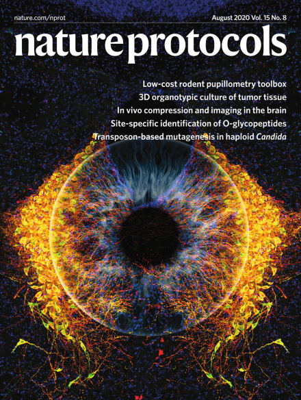
Pupillometry as a readout of locus coeruleus activation
Merged image of a human iris (white) and noradrenergic neurons (yellow) of the locus coeruleus from a DBH-iCre mouse. Visualization of the noradrenergic neurons is a consequence of triple-labeling for tyrosine hydroxylase (green), virally encoded floxed mCherry (red, delivered via an AAV-5 stereotactic injection) and a neuronal marker (Nissl bodies, blue).
Image: Microscopy image taken by Amalia Floriou-Servou, photograph of the iris by Edouard Janssens, image merging by Sarah Steinbacher. Cover Design: Marina Spence.
Here the authors describe an updated protocol for single-stranded sequencing library preparation suitable for highly degraded DNA from ancient remains or other sources. The procedure can be performed manually or in an automated fashion.
Pupil diameter is measured in rodents using low-cost cameras and a Raspberry Pi computer to obtain a rapid, noninvasive real-time readout of locus coeruleus activity.
This protocol describes how to build and implant an in vivo compression device that simulates the solid mechanical forces exerted on the mouse brain by a growing tumor. Standard transparent cranial windows are adapted to include a turnable screw for controlled compression/decompression.
This protocol addresses the challenge of detecting and quantifying known protein complexes at the proteome level using SEC, DIA/SWATH mass spectrometry and a computational framework, CCprofiler.
This protocol describes a lentivirus-based massively parallel reporter assay (lentiMPRA) for quantitative assessment of the activity of gene regulatory elements and an accompanying Nextflow-based computational pipeline for analysis.
This protocol describes the isolation and 3D culture of organoids from fresh murine or human primary and metastatic tumor tissue and provides instructions for real-time imaging, genetic and microenvironmental manipulation and molecular analysis.
This protocol describes an approach for quantitative analysis of membrane deformations by purified proteins at a single-molecule level. Changes in luminal conductance of ultra-short (80- to 400-nm) lipid nanotubes are used as real-time reporters.
This protocol offers a CRISPR-based toolkit, including several variants of ‘classic’ CRISPR–Cas9, along with CRISPRi and CRISPR-base editing systems (CRISPR-BEST) for genome editing in streptomycetes.
This protocol describes a ChIP-seq procedure optimized for profiling H3K27 acetylation in archived formalin-fixed, paraffin-embedded (FFPE) tissues sampled through whole or macrodissected sectioning or from punched cores.
In pseudotargeted metabolomics, transitions used for multiple-reaction monitoring are chosen from mass spectrometry data obtained using mixtures of real samples. Semiquantitative information is obtained without knowing the identity of the compounds.
Nicholson et al. describe a system for identifying molecular species derived from nuclear magnetic resonance spectroscopy-based metabolic phenotyping studies, with detailed information on sample preparation, data acquisition and modeling. They recommend eight modular workflows to be followed in sequential order.
This protocol describes procedures for isolating the protein-bound transcriptome and RNA-binding proteome via UV crosslinking and organic/aqueous phase separation.
This protocol describes a workflow for site-specific extraction and mass spectrometry-based identification of O-linked glycopeptides (EXoO). The protocol can be applied to cultured cells, tissues and plasma.
This protocol describes how to perform geometric morphometrics on microscopic animals, as exemplified by analysis of nematode mouth morphology. The procedure provides guidance for microscopy data acquisition, data analysis and validation.
Treatment with the TLR7/8 ligand (R848) precipitates X sperm, thus facilitating the separation of X sperm from Y sperm. The provided protocols for mouse and bovine sperm separation can then be used in IVF.
This protocol describes nanoflow liquid chromatography–mass spectrometry (nanoLC-MS) analysis of the N-glycans and O-glycans of glycoproteins and glycolipids, as well as site-specific glycosylation of membrane proteins.
This protocol describes a PB transposon-based mutagenesis system for construction of high-quality mutant libraries in haploid Candida species. The procedure describes mutant library generation, PB insertion-site sequencing and data analysis.
In this protocol, the authors describe the design, fabrication, purification, characterization and potential biomedical applications of a self-assembling TDN-based multifunctional delivery system.