
Volume 14 Issue 5, May 2019
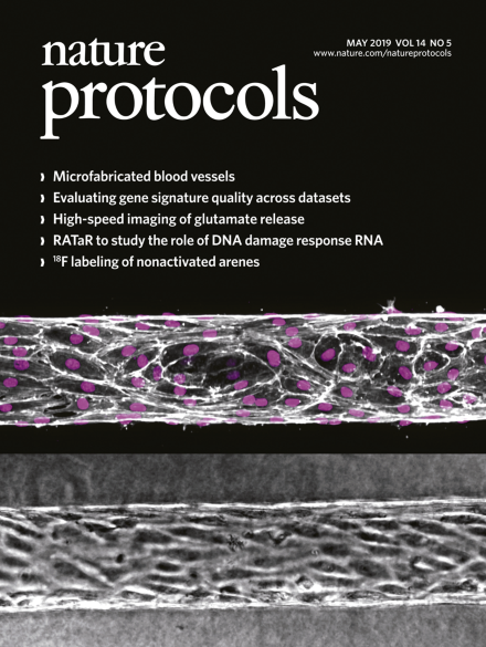
Microfabricated blood vessels
Phase contrast (left) and immunostained micrographs (right) of perfusable human engineered microvessels used to investigate vascular barrier function. Immunostaining for VE-cadherin (white) and nuclei (magenta) demonstrates the formation of endothelial cell–cell adherens junctions that regulate the permeability of the vessel in response to flow.
See Polacheck et al.
Image: William J. Polacheck. Cover design: Erin Dewalt.
Protocols
-
Advertisement
