Thank you for visiting nature.com. You are using a browser version with limited support for CSS. To obtain
the best experience, we recommend you use a more up to date browser (or turn off compatibility mode in
Internet Explorer). In the meantime, to ensure continued support, we are displaying the site without styles
and JavaScript.
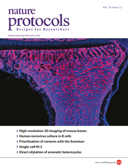
A tile scan confocal microscopic image of a tibial longitudinal section showing the organization of blood vessels in a mouse long bone. Immunofluorescence staining for the endothelial marker endomucin is shown in red. Fluorescently stained nuclei are shown in blue (DAPI). Taken from the protocol by Kusumbe et al. doi:10.1038/nprot.2015.125. Cover design by Jamel Wooten.
Carbon-hydrogen bond functionalization is a desirable strategy for the selective derivatization of organic molecules. This protocol describes the direct silylation of aromatic heterocycles using an inexpensive and abundant catalyst: potassium tert-butoxide.
This protocol describes how to prepare thick sections of mouse skeletal tissue for high-resolution 3D confocal imaging. Samples prepared in this way retain both fluorescence activity and antigenicity, allowing simultaneous imaging of multiple markers.
Targeted RNA directional sequencing (TARDIS) combines targeted RNA capture (using DNA traps) with strand-specific sequencing to enable discovery of rare transcripts, including long and short noncoding RNAs.
Human noroviruses are a leading cause of foodborne disease. Here the authors facilitate the study of these viruses with protocols for culturing them in human B cells and assessing bacterial stimulation of B cell infection and viral attachment to B cells.
Imaging FCS and FCCS generate high-resolution quantitative data over large numbers of pixels that can be used to measure a range of molecular characteristics, including concentration, diffusion rates and interactions.
This protocol describes a platform for directly converting human mid-gestation lineage-committed amniotic fluid-derived cells into a stable and expandable population of vascular endothelial cells over a 3-week period without using pluripotency factors.
The Fraser laboratory provides a protocol for performing Hi-C on single cells. The technique is an important step towards understanding cellular variability in genome organization and chromosome conformation.
This protocol describes use of the Exomiser suite, a collection of algorithms that allow for prioritization of genes and variants from exome sequencing data for disease-gene discovery.
Having demonstrated that brain endothelial cells cross-present parasite antigen in mouse experimental cerebral malaria, the authors present ex vivo and in vitro protocols to measure this antigen presentation, generalizable to other disease contexts.
This protocol uses micropatterned co-cultures comprising 2D islands of primary human hepatocytes surrounded by supportive fibroblast cells to model liver infection by the hepatitis B and C viruses or Plasmodium pathogens in vitro.
Live imaging of regeneration of adult zebrafish tissues has been hampered by the inability to continuously anaesthetize fish for longer than 2 h. Xu et al. describe an intubation-based approach that extends that period to 2 d.
It is important to check whether an expressed membrane protein is successfully secreted to the cell surface. Extracellular protease digestion of intact cells followed by immunoblotting is easy and informative, but it requires an often-omitted control.