Thank you for visiting nature.com. You are using a browser version with limited support for CSS. To obtain
the best experience, we recommend you use a more up to date browser (or turn off compatibility mode in
Internet Explorer). In the meantime, to ensure continued support, we are displaying the site without styles
and JavaScript.
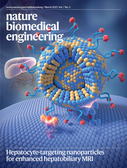
Hepatocyte-targeting nanoparticles for enhanced hepatobiliary MRI
This issue highlights that hepatobiliary MRI can be enhanced by an ultrasmall nanoparticle targeting hepatocytes, that transmembrane water-efflux rate is a sensitive biomarker of the expression of aquaporin-4, a hydrogel-based metamaterial for profiling extracellular vesicles in patient samples, a near-infrared fluorophore for the imaging of aggregates of amyloid-β and tau through the skull of mice, nanoparticles producing ultrasound-induced afterglow luminescence, a library of renally cleared fluorescence probes for the tracking of tumour-infiltrating leukocytes, liposomal nanoparticles for spatially mapping light in deep tissue via MRI, and a tomographic method for the location of photon pairs produced from high-energy X-rays.
The cover illustrates an ultrasmall nanoparticle with a manganese ferrite core and with surface ligands with high specificity for hepatocytes, for use as a contrast agent for imaging the liver and the biliary tree.
Water exchange through the transmembrane channel aquaporin-4 can be measured by conventional dynamic-contrast-enhanced magnetic resonance imaging and is a sensitive biomarker of the proliferation of gliomas and their resistance to chemotherapy.
Liposomal nanoparticles incorporating photosensitive lipids and enclosing paramagnetic molecules enable the mapping, via magnetic resonance imaging, of spatial variations of light intensity in illuminated brain tissue in living animals.
This Perspective discusses the applicability of integrated whole-body PET/MRI for the study of immune-mediated phenomena associated with haematopoietic activity and cardiovascular disease.
Hepatobiliary MRI can be enhanced by an ultrasmall nanoparticle composed of a manganese ferrite core and surface ligands binding to hepatocyte-specific transmembrane metal and anion transporters, as shown in small and large animals.
Transmembrane water-efflux rate is a sensitive biomarker of the expression of aquaporin-4, and can be measured via conventional dynamic-contrast-enhanced magnetic resonance imaging, as shown in animals and in patients with gliomas.
Neuroectodermal cells produced via the direct reprogramming of human astrocytes can be induced to form spinal-cord organoids with functional neurons specific to the dorsal and ventral domains.
A fluorophore with peak excitation and emission wavelengths in the near-infrared window allows for the microscopic imaging of amyloid-β and tau aggregates through the intact skull in mouse models of Alzheimer’s disease.
A library of renally cleared fluorescence probes that are activated only in the presence of both tumour and leukocyte biomarkers allows for the tracking of specific populations of tumour-infiltrating leukocytes in vivo and in urine.
Nanoparticles producing ultrasound-induced afterglow luminescence in the presence of a tumour biomarker allow for the image-guided treatment of mice bearing subcutaneous tumours.
Light in deep tissue can be spatially mapped via magnetic resonance imaging by leveraging liposomal nanoparticles incorporating photosensitive lipids and enclosing paramagnetic molecules.
A tomographic method for the location of photon pairs originating from the annihilation of positron–electron pairs produced by high-energy X-rays in tissue could allow for the monitoring of radiotherapy dosing.