
Volume 90 Issue 6, June 2010
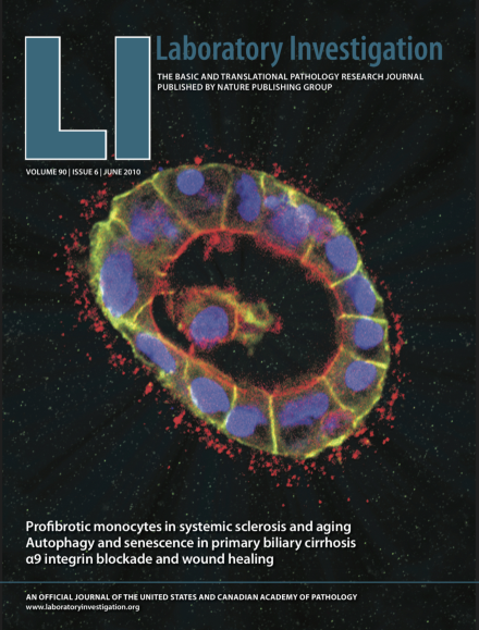
Canine kidney epithelial MDCK cells form a spherical cyst with a lumen inside when cultivated in three dimensional mixture of basement membrane proteins. The cover shows a confocal image of MDCK cell cyst where E-cadherin shown in green delineates the lateral membranes and actin shown in red is concentrated in apical microvilli facing the lumen. The nuclei are shown in blue. The lumen formation requires apoptosis of inner cells and some remnants are still seen within the lumen. For more information see the paper by Töyli et al on page 915.
Inside Lab Invest
-
Advertisement
