Volume 90
-
No. 12 December 2010
Special Issue – Hepatic and Pancreatic SystemsThe cover shows bioimaging of mouse liver oxidative stress after partial hepatectomy. roGFP (reduction/oxidation-sensitive green fluorescent protein) allows real-time visualization of the oxidation/reduction potentials of the organ. roGFP was adenovirally transducted to mouse liver prior to partial hepatectomy. Light emission from redox-sensitive GFP (roGFP) from remnant liver lobes was directly measured and imaged from the exposed liver surface after partial hepatectomy. For more information see the paper by Haga et al, on page 1718
-
No. 10 October 2010
Tartrate-resistant acid phosphatase activity in a culture of osteoclasts. Variable numbers of nuclei, which are unstained, are seen in the centers of cells in negative contrast. The cells were differentiated 10 days in vitro from CD14 purified human peripheral blood mononuclear cells, in DMEM with 5% heat inactivated fetal calf serum with 10 ng/ml of recombinant CSF-1 and 20 ng/ml of recombinant soluble RANKL added. The brightfield photograph is 880 mm, top to bottom. For more information see the paper by Yaroslavskiy et al on page 1533.
-
No. 9 September 2010
TNF-α is a key regulator of adipose tissue mass, but mechanisms underlying this effect have not been fully elucidated. In this issue (page 1385), the paper by Tamai et al shows that TNF-α selectively deletes adipocytes, but not preadipocytes, via induction of apoptosis. The cover shows phase-contrast microscopy of the 3T3-L1 mature adipocytes used in this study.
-
No. 8 August 2010
The cover shows actin polymerization in rat gastric epithelial cells. The photo is adapted from a study by Chai et al (page 1140, this issue), which reveals that matricellular protein CCN1 can induce reversible epithelial-mesenchymal transition and may therefore have clinical utility for wound healing.
-
No. 7 July 2010
Cervical cancer is a major public health problem, and cervical cytology screening requires considerable human effort to screen the millions of pap smears taken each year. Infrared spectroscopy offers an alternative to conventional cytology, whereby infrared micro-spectroscopy can be performed on conventional cytological preparations and coupled with sophisticated data analysis to determine whether or not a cytological sample is abnormal. The cover is an artistic interpretation related to the proof-of-concept paper by Schubert et al. on page 1068–1077.
-
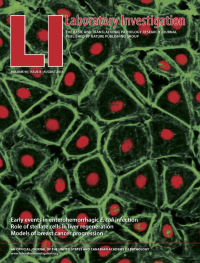
No. 6 June 2010
Canine kidney epithelial MDCK cells form a spherical cyst with a lumen inside when cultivated in three dimensional mixture of basement membrane proteins. The cover shows a confocal image of MDCK cell cyst where E-cadherin shown in green delineates the lateral membranes and actin shown in red is concentrated in apical microvilli facing the lumen. The nuclei are shown in blue. The lumen formation requires apoptosis of inner cells and some remnants are still seen within the lumen. For more information see the paper by Töyli et al on page 915.
-
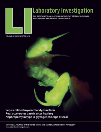
No. 4 April 2010
Live Beta1glo founder lines show strong GFP expression that will subsequently be lost wherever Cre recombinase is expressed to rearrange the transgene and cause conditional overexpression of transforming growth factor-beta 1. For more information see the paper by Hall et al on page 543.
-
No. 3 March 2010
Cover: The paper by Zhang et al in this issue on p 436 shows that taxol treatment suppresses fibroncetin expression in a rat model of unilateral ureteral obstruction. The cover shows a representative sham group rat kidney section subjected to fibronectin immunofluorescence
-
No. 2 February 2010
Cover: Pellet of synovial mesenchymal stem cells during in vitro chondrogenesis. Optical micrograph of pellet stained with toluidine blue is shown. At day 7, the structure of the pellet was divided into three layers. The superficial zone consisted of spindle-shaped cells organized along the surface of the pellet. The deep zone was composed of unorganized polygonal cells with intercellular space. The middle zone could be distinguished between the superficial and the deep zone. For more information see the paper by Ichinose et al on page 210.
Supplement
-
No. 1 January 2010
Cover: The cover shows intravital epi-fluorescence microscopy displaying arteriolo-venular bundles in mice administered erythtopoietin and/or Nω-nitro-L-arginine methylester in a dorsal skinfold chamber. Contrast was enhanced with FITCdextran 150,000, with magnification x80. For more information see the paper by Rezaeian et al on page 40.