


« Prev Next »
Stealthy Skin of Coleoid Cephalopods
The ocean can be a dangerous place, and being a squishy piece of delicious, nutritious muscle is not ideal given that so many capable predators abound. Coleoid cephalopods, a group that includes octopuses, cuttlefish and squid, experience the selective pressure of predation from eels, nurse sharks, and a great many fishes (Aronson, 1991). Yet based on molecular findings, coleoid cephalopods have been present since the early Devonian period, diverging from their ancestor over 400 million years ago (Bergmann et al., 2006). Survival might be hopeless for soft bodied coleoid cephalopods if it were not for camouflage. In addition to hiding in crevices and small holes that these soft-body mollusks easily fit into (Sheel & Bisson, 2012), many cephalopods rely on sophisticated tissues - the chromatophores, iridophores, leucophores and papillae - to blend in with their surroundings and disrupt their body outlines, making them much more difficult to locate by sight. Many coleoids share these tissues and organs, but the common and mimic octopuses (Octopus vulgaris and Thaumoctopus mimicus, respectively) have received much attention in popular media over the past decade (Figure 1).
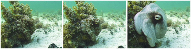
Figure 1
A common octopus (Octopus vulgaris) changes both color and texture after being approached by Dr. Roger Hanlon (used with permission). Full video available at http://www.mbl.edu/mrc/hanlon/video.html
© 2016 Current Biology Courtesy of Roger Hanlon, Marine Biological Laboratory All rights reserved. 
What are chromatophores?
Chromatophores are organs that are present in the skin of many cephalopods, such as squids, cuttlefish, and octopuses, which contain pigment sacs that become more visible as small radial muscles pull the sac open making the pigment expand under the skin. Electrical activity within a chromatophore nerve (Fig. 2, G) causes the radial muscle fibers of the chromatophore (Fig. 2, D) to pull outward toward the perimeter of the chromatophore, expanding the central pigment sack (Fig. 2, A). Early morphologic and physiologic work by Florey (1969) showed that the radial muscles widen the pigment sac with increasing frequency of the nerve electrical activity. The radial muscles are thought to be connected to each other by gap junctions (Florey, 1969) so that they ‘dilate' the chromatophore in a symmetrical fashion. The elastomeric properties of the membrane around the pigment granules -the cytoeslastic sacculus (Fig. 2, C), is thought to be responsible for contracting the chromatophore after it has opened (Florey, 1969). The chromatophores can be opened quickly because they are controlled neurally: squid, cuttlefish and octopuses can change colors within milliseconds (Hanlon, 2007).

Figure 2
(a) Chromatophores in a portion of squid fin skin (used with permission from George Bell, MBL) (b) Anatomy of a chromatophore. The chromatophores are considered organs because of their combination of all categories of animal tissue into a single functional unit – but there are many hundreds distributed through the skin of most cephalopods. A: Pigment granules, B: Nucleus, C: Cytoelastic sacculus, D: Radial muscle fibers, E: Mitochondria, F: Muscle cell, G: Nerve axon, H: Glial cell, I: Primary infoldings and pouches (Adapted from Cloney & Florey, 1968).
A) Courtesy of George bell, Marine Biological Laboratory, Woods Hole B) Cloney, R. A. and E. Florey. 1968. Ultrastructure of cephalopod chromatophore organs. Zellforsch 89:250‐280.
What are iridophores and how do they act in cephalopod camouflage?
Camouflage using chromatophores is particularly impressive because chromatophore pigments are typically only red, yellow, or brown (Hanlon et al., 2011). Yet there are certainly other colors that need to be mimicked but which cannot be made by combining "pixels" of just those three. However, these three colors are particularly useful at the depths wherein many camouflaging cephalopods live (Bush et al., 2009). Other colors are attainable by using a second layer of structures in the cephalopod skin called iridophores (Cooper & Hanlon, 1986).
Iridophores are stacks of very thin cells that are capable of reflecting light back at different wavelengths (Cloney & Brocco, 1983) and possibly different polarities (Mathger & Hanlon, 2007). Interestingly, the color an iridophore reflects is dependent on the angle from which they are observed (Mathger & Hanlon, 2007). When observed from above, iridophores can appear blue, but when observed at a more oblique angle, they appear to reflect red light. By combining reflection from the iridophores with the correct patterning of chromatophores, the cephalopod can create a very convincing copy of the surrounding conditions. Unlike chromatophores, it remains dubious that iridophores are controlled directly by neural inputs because they respond much more slowly (ca. several seconds to minutes) and thus may be controlled by neurohormones, a diffusible cue, or weak electric coupling to an unidentified intermediary.
How does bumpy skin hide a cephalopod? The business of papillae.
Papillae are sections of the skin that can be deformed in order to change texture, and may work by a hydrostatic mechanism (Allen et al., 2009).

Figure 3
Papillae still contain chromatophores and iridophores found in the skin: they are areas where the skin can deform due to pressure (Allen et al., 2013), thus changing the outline of the animal, or in dramatic cases, its shape.
Allen, J., Bell, G. R. R., Kuzirian, A., & Hanlon, R. (2013), Cuttlefish skin papilla morphology suggests a muscular hydrostatic function for rapid changeability. Journal of Morphology 274: 645–656.
Not only is matching the texture of a substrate important for visual blending, having texture on the skin makes the cephalopod display a less identifiable edge. Many vertebrate predators find their prey by looking for visual edges and breaks in the background (Burr et al., 1989). Although it may be somewhat counterintuitive, cephalopods seem to use visual cues and not tactile cues to determine how the papillae should be expressed (Allen et al., 2009). To evaluate this, investigators placed cuttlefish in a tank with either a smooth pattern without sharp contrast, a slightly contrasting pattern, or a highly contrasting pattern and manipulated the texture of the substrate. Each pattern was presented uncovered or covered by glass to give only visual information but no tactile information (Allen et al., 2009). Papillae expression did not change when tactile information was varied, meaning that the cuttlefish being investigated was likely using visual cues.
Leucophores: Specialized Reflectors
Cuttlefish and octopuses possess an additional type of reflector cell called a leucophore. They are cells that scatter full spectrum light so that they appear white in a similar way that a polar bear's fur appears white. Leucophores will also reflect any filtered light shown on them, for instance, they will reflect green light if green is presented to them (Mathger et al., 2009). Unlike iridophores, leucophores do not change appearance based on the viewing angle (Cloney & Brocco, 1983).The leucophores are thought to affect the intensity of the presented chromatophores by providing a white backdrop, aiding in patterns that disrupts the cuttlefish and octopus body outline (Hanlon & Messenger, 1988). Since the leucophores reflect filtered light as well, they aid in color matching because they will reflect wavelengths of light that are filtered by seawater at lower depths.
Is mimicry the right word?
The most mysterious quality about cephalopod crypsis is that cephalopods are believed to be colorblind (Sereni, 1930). While it is certainly impressive that cephalopods can mimic color incredibly well despite being colorblind in their eyes and being capable of mimicking color when the eyes are removed (Sereni, 1930), perhaps knowing the color of the background is not necessary. Mimicry might not be the right term for this phenomenon. For example, cephalopod camouflage might really be sophisticated color disruption. Coleoid cephalopods could display background color (and texture) information at a chromatic and spatial resolution in excess of predator visual acuity. The cephalopod would appear to be part of the background in this scenario because the predators' visual systems cannot discern a difference, which means the cephalopods do not have to match the background perfectly in order to hide (Chiao et al., 2011). Thus, it is plausible that a cephalopod could mimic how disruptive a background is with visual cues and then modulate how much pigment to display based on the light levels detected either centrally or peripherally. But this would require a peripheral photo-sensor. Mathger et al. (2010) detected opsin in the skin of the cuttlefish that differed from opsin found in the eyes by a single amino acid, meaning there is a possibility that coleoid cephalopods can sense how much ambient light is present across their periphery, and adjust their skin color and brightness accordingly. The underlying iridophores would be able to reflect light to further refine the color of their skin and the papillae would be activated based on the perceived substrate to further disrupt the coleoid cephalopod's body outline. This could be how cephalopods are often able to fool predators and their skin is able to blend with the background with a higher resolution than predators can detect.
Summary
Cephalopod camouflage is among the most dynamic in the animal kingdom, helping their lineage of soft-bodied and otherwise vulnerable relatives survive for hundreds of millions of years. While the individual components of the camouflage system have received extensive study and are relatively well understood, how cephalopods choose which camouflaging patterns to express in different circumstances is still quite mysterious. The different control mechanisms for chromatophores, iridophores, leucophores and papillae require cephalopods to integrate different types of visual information into a cohesive, matching pattern. How their brains process visual information from their eyes and possibly also their skin, then send out the correct commands to their camouflaging tissues, is something we do not yet understand. Knowing more about the stealthy skin tricks of cephalopods can help us understand more about their behavior and evolution, and might also be useful for developing our own camouflaging materials in the future. Maybe one day our nations' soldiers will be wearing camouflaging clothing that matches their background and reduces high contrast silhouette lines as quickly and as well as cephalopod skin does.
Special thanks to Dr. Roger Hanlon & George Bell at The Marine Biological Laboratory
References and Recommended Reading
Allen, J., Bell, G. R. R., Kuzirian, A., & Hanlon, R. (2013), Cuttlefish skin papilla morphology suggests a muscular hydrostatic function for rapid changeability. Journal of Morphology 274: 645-656.
Allen, J., Mathger, L., Barbosa, A., Hanlon, R. (2009). "Cuttlefish use visual cues to control three dimensional skin papillae for camouflage." Journal of Comparative Physiology A 195: 547-555.
Aronson, R. (1991) "Ecology, paleobiology and evolutionary constraint in the octopus." Bulletin of Marine Science 49: 245-255.
Bergmann, S., Lieb, B., Ruth, P., & Markl, J. (2006) "The hemocyanin from a living fossil, the cephalopod Nautilus pompilius: Protein structure, gene organization and evolution." Journal of Molecular Evolution 62: 362-374.
Burr DC, Morrone MC, Spinelli D (1989) Evidence for edge and bar detectors in human vision. Vision Research 29:419-431
Chiao, C., Wickieser, J., Allen, J., Genter, B., Hanlon, R. (2011). "Hyperspectral imaging of cuttlefish camouflage indicates good color match in the eyes of fish predators." Proceedings of the National Academy of Sciences 108: 9148-9153
Cloney, R. & Brocco, S. (1983). "Chromatophore organs, reflector cells, iridocytes, and leucophores." American Zoologist 23: 581-592.
Cloney, R. A. and E. Florey. 1968. Ultrastructure of cephalopod chromatophore organs. Zellforsch 89:250-280.
Cooper, K.M., Hanlon, R. (1986). "Correlation of iridescence with changes in iridophore platelet ultrastructure in the squid lliguncula brevis." J Exp Biol. 121: 451-5.
Florey, E. (1969), "Ultrastructure and function of cephalopod chromatophores." Am Zool. 9:429-442.
Hanlon, R. (2007). "Cephalopod dynamic camouflage." Current Biology 17: 400-404.
Hanlon, R., Chiao C., L. Mathger, K., Buresch, Barbosa,A., Allen., J., Siemann, L. Chubb,C. (2011) "Rapid adaptive camouflage in cephalopods." in Animal Camouflage: Mechanisms and Function. Cambridge University Press, Cambridge, UK. pp145-161.
Hanlon, R. & Messenger, J. (1988). "Adaptive coloration in young cuttlefish (Sepia officianlis L.): The morphology and development of body patterns and their relation to behaviour." Philosophical Transactions of the Royal 320: 437-487.
Mathger, L. & Hanlon, R. (2007). "Anatomical basis for camouflaged polarized light communication in squid." Biological Letters 2: 464-496.
Mathger, L., Denton, E., Marshall, N., Hanlon, R. (2009). "Mechanisms and behavioural functions of structural coloration in cephalopods." Journal of the Royal Society Interface 6: S149-S163.
Sereni, E. "The chromatophores of the cephalopods." Biological Bulletin 59, 3. 247-268.
Sheel, D., & Bisson, L. (2012). "Movement patterns of giant Pacific octopuses, Enteroctopus dofleini." Journal of Experimental Marine Biology and Ecology 416-17: 21-31.
Wardill, T., Gonzalez-Bellido, P., Crook, R., Hanlon, R. (2012). "Neural control of tuneable skin iridescence ins squid." Proceedings of the Royal Society B 279: 4243-4252.


