 that wind together to form a 24 nm wide hollow cylinder. Microtubules are structures that can rapidly grow (via polymerization) or shrink (via depolymerization) in size, depending on how many tubulin molecules they contain.", "true", "All rights reserved.", '655', '527', 'http://www.molbiolcell.org/site/misc/terms.xhtml');)
 that wind together to form a 24 nm wide hollow cylinder. Microtubules are structures that can rapidly grow (via polymerization) or shrink (via depolymerization) in size, depending on how many tubulin molecules they contain.
", "
true
", "
All rights reserved.
", '
655
', '
527
', '
http://www.molbiolcell.org/site/misc/terms.xhtml
');)
 Neurons are specialized eukaryotic cells that extend long processes to form connections in the nervous system. Like other eukaryotic cells, neurons have a cytoskeleton that consists of three main polymers: microtubules (green), intermediate filaments (purple) and actin filaments (red). Microtubules emanate from the axon, and actin-filament networks form sheet-like structures and filopodial protrusions at the leading edge. Scale bar, 20 μm. (C) The neuronal axon is a long membrane-bounded extension, in which neurofilaments (a class of intermediate filament in neurons) form a structural matrix that embeds microtubules, which transport materials from the cell body to the axon terminals at the synapse. (D) The growth cone contains dendritic actin filament networks and parallel actin filament filopodia. (E) Microtubules consist of 13 protofilaments of tubulin dimers arranged in a hollow tube. (F) Neurofilaments have flexible polymer arms that repel neighboring neurofilaments and determine the radius of the axon. (G) Actin filaments are arranged into networks. These networks can have many architectures, including the branched structures depicted here, which are formed by the Arp2/3 complex (blue). The diameters of microtubules, intermediate filaments, and actin filaments are within a factor of three of each other; the diagrams in E, F, and G are drawn approximately to scale. However, the relative flexibilities of these polymers differ markedly, as indicated by their persistence lengths: from least to most flexible, microtubules (5,000 μm), actin filaments (13.5 μm) and intermediate filaments (0.5 μm).", "true", "All rights reserved.", '456', '493', 'http://www.nature.com');)
 Neurons are specialized eukaryotic cells that extend long processes to form connections in the nervous system. Like other eukaryotic cells, neurons have a cytoskeleton that consists of three main polymers: microtubules (green), intermediate filaments (purple) and actin filaments (red). Microtubules emanate from the axon, and actin-filament networks form sheet-like structures and filopodial protrusions at the leading edge. Scale bar, 20 μm. (C) The neuronal axon is a long membrane-bounded extension, in which neurofilaments (a class of intermediate filament in neurons) form a structural matrix that embeds microtubules, which transport materials from the cell body to the axon terminals at the synapse. (D) The growth cone contains dendritic actin filament networks and parallel actin filament filopodia. (E) Microtubules consist of 13 protofilaments of tubulin dimers arranged in a hollow tube. (F) Neurofilaments have flexible polymer arms that repel neighboring neurofilaments and determine the radius of the axon. (G) Actin filaments are arranged into networks. These networks can have many architectures, including the branched structures depicted here, which are formed by the Arp2/3 complex (blue). The diameters of microtubules, intermediate filaments, and actin filaments are within a factor of three of each other; the diagrams in E, F, and G are drawn approximately to scale. However, the relative flexibilities of these polymers differ markedly, as indicated by their persistence lengths: from least to most flexible, microtubules (5,000 μm), actin filaments (13.5 μm) and intermediate filaments (0.5 μm).
", "
true
", "
All rights reserved.
", '
456
', '
493
', '
http://www.nature.com
');)


« Prev Next »

Microtubules and Filaments
The cytoskeleton is a structure that helps cells maintain their shape and internal organization, and it also provides mechanical support that enables cells to carry out essential functions like division and movement. There is no single cytoskeletal component. Rather, several different components work together to form the cytoskeleton.
What Is the Cytoskeleton Made Of?
The
cytoskeleton of eukaryotic cells is made of filamentous proteins, and it
provides mechanical support to the cell and its cytoplasmic
constituents. All
cytoskeletons consist of three major classes of elements that differ in size and in protein composition.
Microtubules are the largest type of filament, with a diameter of about
25
nanometers (nm), and they are composed of a protein called tubulin.
Actin filaments are the smallest type, with a diameter of
only about 6 nm, and they are made of a protein called actin.
Intermediate filaments, as their name suggests, are
mid-sized, with a diameter of about 10 nm. Unlike actin filaments and
microtubules,
intermediate filaments are constructed from a number of different
subunit
proteins.
What Do Microtubules Do?
Tubulin contains two polypeptide subunits, and dimers of these subunits string together to make long strands called protofilaments. Thirteen protofilaments then come together to form the hollow, straw-shaped filaments of microtubules. Microtubules are ever-changing, with reactions constantly adding and subtracting tubulin dimers at both ends of the filament (Figure 1). The rates of change at either end are not balanced — one end grows more rapidly and is called the plus end, whereas the other end is known as the minus end. In cells, the minus ends of microtubules are anchored in structures called microtubule organizing centers (MTOCs). The primary MTOC in a cell is called the centrosome, and it is usually located adjacent to the nucleus.
Microtubules tend to grow out from the centrosome to the plasma membrane. In nondividing cells, microtubule networks radiate out from the centrosome to provide the basic organization of the cytoplasm, including the positioning of organelles.
What Do Actin Filaments Do?
The protein actin is abundant in all eukaryotic cells. It was first discovered in skeletal muscle, where actin filaments slide along filaments of another protein called myosin to make the cells contract. (In nonmuscle cells, actin filaments are less organized and myosin is much less prominent.) Actin filaments are made up of identical actin proteins arranged in a long spiral chain. Like microtubules, actin filaments have plus and minus ends, with more ATP-powered growth occurring at a filament's plus end (Figure 2).
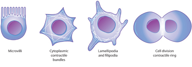
In many types of cells, networks of actin filaments are found beneath the cell cortex, which is the meshwork of membrane-associated proteins that supports and strengthens the plasma membrane. Such networks allow cells to hold — and move — specialized shapes, such as the brush border of microvilli. Actin filaments are also involved in cytokinesis and cell movement (Figure 3).
Figure 3: Actin filaments support a variety of structures in a cell.
© 2014 Nature Education All rights reserved. 
What Do Intermediate Filaments Do?
Intermediate filaments come in several types, but they are generally strong and ropelike. Their functions are primarily mechanical and, as a class, intermediate filaments are less dynamic than actin filaments or microtubules. Intermediate filaments commonly work in tandem with microtubules, providing strength and support for the fragile tubulin structures.
All cells have intermediate filaments, but the protein subunits of these structures vary. Some cells have multiple types of intermediate filaments, and some intermediate filaments are associated with specific cell types. For example, neurofilaments are found specifically in neurons (most prominently in the long axons of these cells), desmin filaments are found specifically in muscle cells, and keratins are found specifically in epithelial cells. Other intermediate filaments are distributed more widely. For example, vimentin filaments are found in a broad range of cell types and frequently colocalize with microtubules. Similarly, lamins are found in all cell types, where they form a meshwork that reinforces the inside of the nuclear membrane. Note that intermediate filaments are not polar in the way that actin or tubulin are (Figure 4).
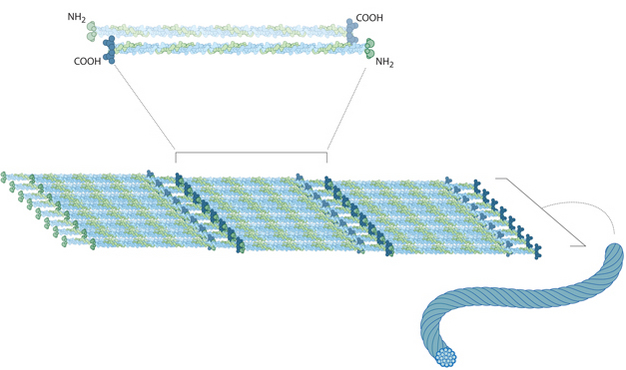
Figure 4: The structure of intermediate filaments
Intermediate filaments are composed of smaller strands in the shape of rods. Eight rods are aligned in a staggered array with another eight rods, and these components all twist together to form the rope-like conformation of an intermediate filament.
© 2014 Nature Education All rights reserved.
How Do Cells Move?
Cytoskeletal filaments provide the basis for cell movement. For instance, cilia and (eukaryotic) flagella move as a result of microtubules sliding along each other. In fact, cross sections of these tail-like cellular extensions show organized arrays of microtubules.
Other cell movements, such as the pinching off of the cell membrane in the final step of cell division (also known as cytokinesis) are produced by the contractile capacity of actin filament networks. Actin filaments are extremely dynamic and can rapidly form and disassemble. In fact, this dynamic action underlies the crawling behavior of cells such as amoebae. At the leading edge of a moving cell, actin filaments are rapidly polymerizing; at its rear edge, they are quickly depolymerizing (Figure 5). A large number of other proteins participate in actin assembly and disassembly as well.
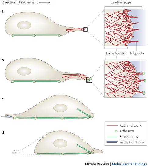
Figure 5: Cell migration is dependent on different actin filament structures.
(A) In a cell, motility is initiated by an actin-dependent protrusion of the cell’s leading edge, which is composed of armlike structures called lamellipodia and filopodia. These protrusive structures contain actin filaments, with elongating barbed ends orientated toward the plasma membrane. (B) During cellular arm extension, the plasma membrane sticks to the surface at the leading edge. (C) Next, the nucleus and the cell body are pushed forward through intracellular contraction forces mediated by stress fibers. (D) Then, retraction fibers pull the rear of the cell forward.
© 2008 Nature Publishing Group Mattila, P.K. & Lappalainen, P. Filopodia: molecular architecture and cellular functions. Nature Reviews Molecular Cell Biology 9, 446-454 (2008). All rights reserved.
Conclusion
The cytoskeleton of a cell is made up of microtubules, actin filaments, and
intermediate filaments. These structures give the cell its shape and
help
organize the cell's parts. In addition, they provide a basis for
movement and
cell division.
eBooks
This page appears in the following eBook


 that wind together to form a 24 nm wide hollow cylinder. Microtubules are structures that can rapidly grow (via polymerization) or shrink (via depolymerization) in size, depending on how many tubulin molecules they contain.
", "
true
", "
All rights reserved.
", '
655
', '
527
', '
http://www.molbiolcell.org/site/misc/terms.xhtml
');)
 Neurons are specialized eukaryotic cells that extend long processes to form connections in the nervous system. Like other eukaryotic cells, neurons have a cytoskeleton that consists of three main polymers: microtubules (green), intermediate filaments (purple) and actin filaments (red). Microtubules emanate from the axon, and actin-filament networks form sheet-like structures and filopodial protrusions at the leading edge. Scale bar, 20 μm. (C) The neuronal axon is a long membrane-bounded extension, in which neurofilaments (a class of intermediate filament in neurons) form a structural matrix that embeds microtubules, which transport materials from the cell body to the axon terminals at the synapse. (D) The growth cone contains dendritic actin filament networks and parallel actin filament filopodia. (E) Microtubules consist of 13 protofilaments of tubulin dimers arranged in a hollow tube. (F) Neurofilaments have flexible polymer arms that repel neighboring neurofilaments and determine the radius of the axon. (G) Actin filaments are arranged into networks. These networks can have many architectures, including the branched structures depicted here, which are formed by the Arp2/3 complex (blue). The diameters of microtubules, intermediate filaments, and actin filaments are within a factor of three of each other; the diagrams in E, F, and G are drawn approximately to scale. However, the relative flexibilities of these polymers differ markedly, as indicated by their persistence lengths: from least to most flexible, microtubules (5,000 μm), actin filaments (13.5 μm) and intermediate filaments (0.5 μm).
", "
true
", "
All rights reserved.
", '
456
', '
493
', '
http://www.nature.com
');)