Thank you for visiting nature.com. You are using a browser version with limited support for CSS. To obtain
the best experience, we recommend you use a more up to date browser (or turn off compatibility mode in
Internet Explorer). In the meantime, to ensure continued support, we are displaying the site without styles
and JavaScript.
Label-free X-ray microscopy of nanomedicines and organelles in intact single cells at nanometer resolution using synchrotron radiation
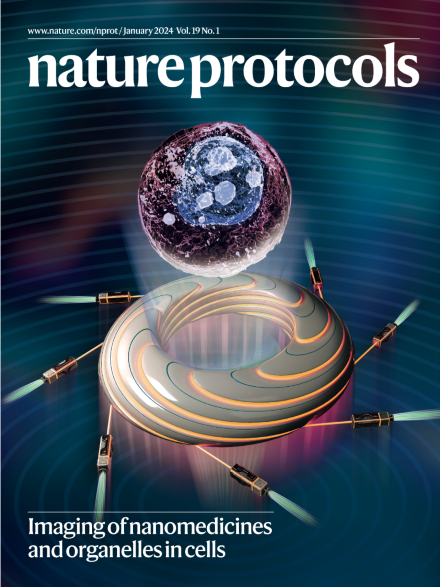
A composite image of a single cell visualized in 3D and a synchrotron radiation facility. The method uses X-rays generated via synchrotron radiation and enables the subcellular localization of nanomedicines in single cells, at nanometer resolution, as a robust approach to characterize interactions between nanomaterials and cells. See Cao et al.
Image: Mingjing Cao, Yaling Wang and Chunying Chen, National Center for Nanoscience and Technology of China. Cover design: S. Whitham.
In 2023, Nature Protocols launched four new collections (on organoids, neural probes, cognitive neuroscience frameworks and neurostimulation) to help readers navigate our content and highlight areas of particular current interest.
This protocol enables genetic manipulation of nuclear and cytoplasmic giant viruses and their host, Acanthomoeba castellanii, by using either CRISPR–Cas9 or homologous recombination.
An X-ray microscopy approach for 2D or 3D label-free visualization of the intracellular distribution of nanomedicines and the morphology of organelles in primary blood cells, macrophages, dendritic cells, monocytes and cancer cells.
This protocol presents a computational approach to scoring drug sensitivity that integrates multiple dose–response parameters into a single response metric and identifies differences in drug-response patterns between cancer cells and healthy control cells.
This protocol provides guidelines for performing single-cell combinatorial indexing cleavage under targets and tagmentation. This method builds on the existing cleavage under targets and tagmentation method and uses a combinatorial indexing step to allow single-cell profiling of chromatin modifications.
This protocol describes an assay to control mechanical forces during the natural flexion-extension cycle of the femorotibial joint of adult living fruit flies and to monitor fluorescent mechanosensitive reporters expressed in the neurons responsible for mechanosensation
Structural and functional analysis of human proteins requires their isolation in high yields and purity. Here, protease-cleavable, high-affinity, tag-specific nanobodies are used for the isolation of soluble and membrane proteins from lentiviral-transduced human cell suspensions.
CONIPHER is a computational framework for accurately inferring subclonal structure and the phylogenetic tree from multisample tumor sequencing, accounting for both copy number alterations and mutation errors.
This protocol describes a reproducible and reliable method for excisional skin wound healing assays in mice. The use of lineage-tracing assays to investigate the contribution of different cell populations to the repair process is also discussed.
This protocol describes barcoding bacteria for identification and quantification, a method for identifying and quantifying bacterial cells in microbiota samples based on the droplet-based barcoding and amplification of 16S rRNA genes from single bacterial cells.
Angstrom-scale two-dimensional channel devices can be assembled with precise control over their dimensions, as a single channel or hundreds of channels using layered crystals, and enable the measurement of angstrom-scale gas, ion and water fluidics.