Thank you for visiting nature.com. You are using a browser version with limited support for CSS. To obtain
the best experience, we recommend you use a more up to date browser (or turn off compatibility mode in
Internet Explorer). In the meantime, to ensure continued support, we are displaying the site without styles
and JavaScript.
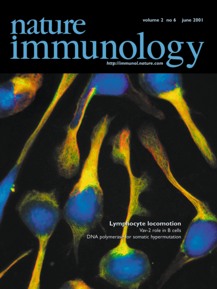
The binding of LFA-1 on a T cell's surface is a critical step for intravascular adhesion and cell migration. Volkov et al. report that, in the T cell's uropod, LFA-1 activates the PKC-(I) (green) pathway to trigger the polymer-ization of tubulin (red) and there-by initiate the migratory capacity of the cells. Blue, nuclei; yellow-orange, colocalization of PKC-(I) and tubulin. Fluorescent image taken by Y. Volkov with an 100x oil immersion lens on a Nikon TE300 microscope and Photometrics-cooled CCD camera.
The events that enable polarization of T cells and migration across the vasculature to the target are largely unknown. New evidence suggests that PKC-β(I) may be pivotal in controlling this process.
Our current understanding of lymphocyte migration across the endothelium includes four steps: attachment, rolling, arrest and diapedesis. New evidence suggests the involvement of another step, chemorheotaxis.
T cells, move over. B cells are now reported, in a recent paper in Nature, to have their own immunological synapses with APCs. Only this time the antigen is whole and B cells don't keep it to themselves.
Genetic ablation experiments have shown that Vav is critical for TCR signaling. Evidence is now emerging that the Vav family of signaling molecules play a critical role in antigen receptor signaling in B cells as well as in T cells.
Multiple error-prone DNA polymerase appear to contribute to immunoglobulin somatic hypermutation. Genetic and biochemical data now indicate that DNA polymerase η may be responsible for the generation of some of the strand-biased hotspot A mutations in the immunoglobulin loci.