Volume 2
-
No. 12 December 2001
Sweat glands are thought of primarily as organs for temperature regulation. Schittek et al. on page 1133 found another valuable function in human sweat: a novel antimicrobial peptide, which they called dermicidin (red). Myoepithelial cells (blue) surround the secretory coil of the eccrine sweat glands (nuclei are green). Dermcidin had antimicro-bial activity at pH and salt concentrations of sweat, making it a candidate for the skin's innate defenses.
-
No. 11 November 2001
Artist's rendition of LCMV-specific memory CD8 + T cells (yellow) in the lung. Selin and colleagues show reactivation of CD8 + T cells (green) specific for LCMV (red) after acute infection with vaccinia virus correlated with a more rapid clearance of vaccinia virus from the mice, decreased mortality and dramatic changes in pathology. (See also News & Views by Phillips, page 991.) Painting in oil on canvas by Mark Sokoloff.
-
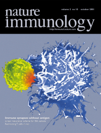
No. 10 October 2001
Scanning electron micrograph of a 7 μm T cell (yellow) interacting with a dendritic cell (blue). Contact through the immune synapse initiates the transmission of signals (symbolized by red) within the T cell from the site of contact. Trautman et al. and Martin et al. have examined the fate of T cells that interact with dendritic cells in the absence of antigen. Micrograph prepared with technical assistance of M. Grasset.
-
No. 9 September 2001
Red plaster mask representing the twisting of the immune system, normally a protector of health and well being (right side of mask) into an instrument of destruction and dysregulation (left side). Half of this month's issue (pages 755 to 822) is focused on the new theoretical frameworks that have arisen to explain autoimmunity and recent developments in treatment of autoimmune disorders. Additional features are available free on Nature Immunology's website (immunol.nature.com). Mask by Christopher Cassidy.
Focus
-
No. 8 August 2001
Artistic interpretation of rheumatoid arthritis synovium (black). A recent mouse model of arthritis highlighted glucose-6-phosphate isomerase (GPI) as a target for both autoreactive antibodies and T cells. Now Ditzel and colleagues show that GPI (green in the tissue section) is a target of antibodies in the human disease, as well. Fluorescence micrograph of a synovium from article on page 746. Red denotes propridium iodide–stained nuclei. Painting is acrylic on paper by Michael Malicki.
-
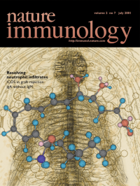
No. 7 July 2001
Resolution of inflammatory infiltrates is intricately regulated by many factors. Serhan and colleagues (page 612) now show that, over time, the production of lipoxygenase products switches to those that inhibit the infiltration of neutrophils from the vasculature, such as lipoxin A4 (a stable analog of which is on the cover). LXA4 is a product of 15-lipoxygenase, which is induced by prostaglandin E2 late in the response.
-
No. 6 June 2001
The binding of LFA-1 on a T cell's surface is a critical step for intravascular adhesion and cell migration. Volkov et al. report that, in the T cell's uropod, LFA-1 activates the PKC-(I) (green) pathway to trigger the polymer-ization of tubulin (red) and there-by initiate the migratory capacity of the cells. Blue, nuclei; yellow-orange, colocalization of PKC-(I) and tubulin. Fluorescent image taken by Y. Volkov with an 100x oil immersion lens on a Nikon TE300 microscope and Photometrics-cooled CCD camera.
-
No. 5 May 2001
Inhibitory NK cell receptors are usually thought of as binding to MHC class I–like proteins. On page 436 of this issue Castells et al. report that a widely expressed integrin can bind to NK receptors on mast cells and inhibit secretion.This could be an effective mechanism for preventing unprovoked mast cell degranulation in the host. αvβ3-expressing T cells (red) binding to gp49B1-expressing cells (green) from fluorescence micrograph in Castells et al.
-
No. 4 April 2001
The epithelial and M cells of the intestine are credited with protecting the host from pathogen invasion through the gastrointestinal tract. Rescigno et al. report on page 361 that dendritic cells can form tight junctions and intercalate between epithelial cells, a location from which they are well positioned to contribute to host defense. See also the News & Views by Gewirtz and Madara on page 288. Welded steel relief of dendritic cells by Christopher Cassidy; original micrograph from Rescigno et al.
-
No. 3 March 2001
horror autotoxicusmastzellenet alpage 216page 193Background: photomicrograph (magnification x300) is extensive vascular congestion in liver after challenge with same peptide.
-
No. 2 February 2001
et al.Science289pages 89pages 91pages 93pages 95pages 102pages 108pages 116pages 123pages 129–134Our Chemokines website (ni/archive/suppinfo/index.html) is free and contains even more features.
Focus
-
No. 1 January 2001
Neo-expressionistic depiction, inspired by A. Kiefer (b. 1945, Donaueschingen, Germany), of the massive deposition of complement (reddish-brown) at the maternal–fetal interface of an embryo, 9.5 days post coitum, in the absence of tryptophan catabolism. Mellor et al. (page 64) show that this deposition is antigen-driven, T cell-dependent and can occur in the complete absence of B cells. Original immunohistochemistry by Mellor et al.