
Volume 6 Issue 6, June 2011
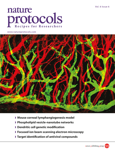
A confocal image of FGF-2induced corneal new blood vessels (CD31, red) and lymphatic vessels (LYVE-1, green). Corneal lymphatic networks comprise large-diameter vessels with blunt-ended vascular sprouts, whereas blood vessel networks are composed of smaller microvessels at higher densities relative to lymphatics.
Protocol
-
Advertisement
