Thank you for visiting nature.com. You are using a browser version with limited support for CSS. To obtain
the best experience, we recommend you use a more up to date browser (or turn off compatibility mode in
Internet Explorer). In the meantime, to ensure continued support, we are displaying the site without styles
and JavaScript.
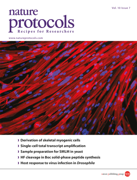
Shown are multinucleated skeletal myotubes derived from human induced pluripotent stem cells generated from a person with Duchenne muscular dystrophy and assessed by immunofluorescence staining for myosin heavy chain (red). Nuclei are shown in blue. Taken from the protocol by Maffioletti et al. doi: 10.1038/nprot.2015.057. Cover design by Jamel Wooten.
In this protocol, pluripotent stem cells are first differentiated into expandable cells resembling human mesoangioblasts; subsequently, transient MyoD induction drives differentiation into multinucleated myotubes.
A common set of reprogramming factors is used to temporarily activate somatic cells into a transition state; these cells are subsequently directed to various cell types via lineage-specific signals, circumventing the need to establish a pluripotent state.
This protocol enables amplification of the total transcript of a single prokaryotic cell for in-depth analysis. Laser-capture microdissection is used to isolate single cells, and amplified cDNA can be further analyzed by microarray.
Human neural progenitor cells with familial Alzheimer's disease mutations are differentiated in a 3D Matrigel matrix, leading to the extracellular aggregation of amyloid-β peptides and accumulation of hyperphosphorylated and aggregated tau protein.
Preserving cell structure and protein localization during sample preparation is crucial for accurate superresolution microscopy. This protocol uses cell wall-penetrating labeling agents, enabling single-molecule localization microscopy in yeast with intact cell walls.
Protein S-sulfenylation is a reversible oxidative modification of cysteine thiol groups. Modified sites react with a dimedone-based probe, DYn-2 which can be used for both enrichment and detection by mass spectrometry as described in this protocol.
This protocol describes a surgical tooth root fenestration model for studying novel approaches and materials used for the regeneration of multi-tissue bone–ligament interfaces in rodents.
Structure-seq applies chemically based RNA structure probing on a genome-wide scale, allowing the in vivo structures of thousands of distinct transcripts to be discerned, and the meta-properties of RNA structure–function relationships to be unveiled.
In Boc-mediated solid-phase peptide synthesis, HF is used to remove protecting groups and to release the peptide from the resin. This protocol provides advice on general methodology, use of cleavage apparatus and safe handling of HF.
Drosophila is a useful model for identifying and characterizing the genetic components involved in host resistance and tolerance to virus infection. The van Rij laboratory explains how to conduct such experiments, controlling for common confounding factors.
This protocol summarizes the recommendations developed after a comparison of Elispot plate-reading approaches by a panel of laboratories and subsequent agreement regarding the optimal steps to follow when evaluating Elispot plates.
This protocol describes the use of two-photon laser-scanning microscopy (TPLSM) to study hair regeneration within a living, uninjured mouse, enabling time-resolved imaging of single hair follicle stem cells.