Thank you for visiting nature.com. You are using a browser version with limited support for CSS. To obtain
the best experience, we recommend you use a more up to date browser (or turn off compatibility mode in
Internet Explorer). In the meantime, to ensure continued support, we are displaying the site without styles
and JavaScript.
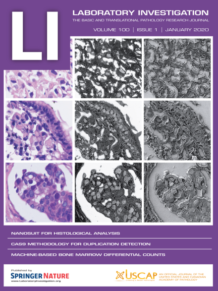
Cover: The cover shows identification of nuclei in paraffin sections using the NanoSuit method. Upper row, malignant mesothelioma; middle row, gastric carcinoma (signet ring cell carcinoma); and lower row, breast cancer sample. Left column, hematoxylin and eosin (H&E) staining; middle column, low-vacuum scanning electron microscopy images of samples stained with gold (III) chloride, taken in BSE mode; and right column, mixed low-vacuum scanning electron microscopy images of samples stained with gold (III) chloride. For more information, see the paper by Kawasaki et al, this issue (p 161).
The authors established and characterized a novel gastroesophageal junction (GEJ) cancer cell line derived from the malignant pleural effusion of a treated GEJ cancer patient. This cell line represents a valuable addition to the limited number of bona fide GEJ cancer cell lines for both basic and translational research.
The severe immunodeficiency of NOD.Cg-PrkdcscidIl2rgtm1Wjl (NSG) mice, a widely used strain for generating patient-derived xenografts from primary tumors, impairs drug metabolism and reduces tolerability towards common cytostatic drugs. Consequently, dose finding and tolerance tests are strongly recommended prior to perform large-scale cancer precision medicine approaches.
We describe generation of neuroblastoma cell lines using conditional reprogramming (CR)—new cell culture method that allows indefinite propagation of cells in an undifferentiated state. We optimized CR growth conditions for neuroblastoma tissue and grew mouse neuroblastoma cells in vitro. These cells demonstrate a unique phenotype, are positive for neuroblastoma markers, and can be passaged and biobanked for continued studies.
This study shows a negative regulation of autophagy-related RNA transcripts and the upregulation and downregulation of p62 and LC3-B proteins, respectively, in the central nervous system of scrapie-infected transgenic mice at clinical stage. These findings likely reflect an impairment of the autophagic pathway at the late symptomatic stage of prion infection.
In chondrocytes, TGF-β1 increases the expression of hypertrophic genes, suggesting that this treatment could mimic osteoarthritis in vitro. EZH2 and Sik3 inhibition, as well as HIF induction repress the expression of hypertrophy markers in TGF-β stimulated chondrocytes, validating the reliability of this model to predict anti-hypertrophic effects of drugs.
The authors examined the capability of propagation-based phase-contrast tomography (PB-PCT) with single-distance phase retrieval for longitudinal in vivo monitoring of bone changes using monochromatic synchrotron light. The application of PB-PCT to the measurement of bone defect healing in mice demonstrated its potential for tracking volumetric and mineral-density changes in newly formed bone.
The authors provide a side-by-side comparison of morphology and function of primary ductal fragments isolated from mouse pancreas and pancreatic organoid cultures. Using state-of-the-art techniques, they demonstrate that pancreatic organoids can be a suitable and robust model to study pancreatic ductal epithelial ion transport in health and disease.
Bone marrow aspirate (BMA) differential cell counts (DCCs) are critical for classification of hematologic disorders. Manual DCCs are still considered the gold standard as a reliable automated method is yet to be developed. Digital pathology and machine learning represent a highly promising technology for this purpose. The authors report their experience developing machine learning algorithms to detect and classify BMA cells. Promising early results signify an important initial step in the effort to devise a reliable, objective method to automate DCCs.
The authors designed an adapter multiplex PCR amplicon with multiple domain melting curves to rapidly detect and genotype at least 20 ALK fusion variants in one tube with HRM. In addition, they introduced feature extraction of the second derivative curve to assist visual analysis to automatically discriminate ALK fusion variants.
Digital image analysis (DIA) of multiplex fluorescence-based immunohistochemistry and visual chromogenic evaluation of CDX2, SOX2, SOX9, E-cadherin, and β-catenin in colorectal cancer are comparable, recognizing prognostic value of CDX2 and negative correlation with SOX2. Membrane staining is best evaluated visually, while DIA enables single-cell coexpression analysis and improves visualization and detection of clinicopathological and biological associations.
The paper describes a fully automated numerical analysis for an unbiased quantification of non-alcoholic fatty liver disease (NAFLD) histological features in rodent models. The technique offers a quantitative high-throughput method to rapidly detect NAFLD in large preclinical studies and for accurately monitoring disease evolution.
The NanoSuit method permits the study of paraffin sections using correlative light and electron microscopy at low and high magnification, with the following features: (i) the integrity of the glass slide is maintained, (ii) 3D microstructures of tissue and pathogens can be visualized, (iii) nuclei and 3,3′-diaminobenzidine-stained areas are distinct because of gold chloride usage, (iv) immunohistochemical staining is quantitative, and (v) contained elements can be analyzed.