
Volume 16 Issue 4, April 2021
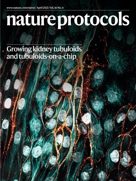
Apical view on a kidney tubuloid cell–derived tubule cultured under perfusion flow in a microfluidic platform (the OrganoPlate).
The image displays immunofluorescence staining of the nuclei (white), sodium-potassium-ATPase (orange) and microtubules (cyan).
See Gijzen et al.
Image: Linda Gijzen and Dorota Kurek, Mimetas BV (imaging); Fjodor A. Yousef Yengej, Hubrecht Institute and University Medical Center Utrecht (editing). Cover design: Tulsi Voralia.
Review Articles
-
Advertisement
