Volume 16
-
No. 12 December 2021
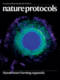
Cardiomyocytes and mesenchymal cells in heart-forming organoidsWhole-mount immunofluorescence staining of a heart-forming organoid showing cardiomyocytes in green, mesenchymal cells in magenta and nuclei in blue (using an anti-myosin heavy-chain antibody, an anti-vimentin antibody and DAPI stain, respectively).
See Drakhlis et al.
-
No. 11 November 2021
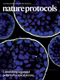
Apical-out human enteroidEnteroids grown in extracellular matrix (ECM) scaffolds develop apical-in polarity, with the microvilli facing the internal lumen. We reversed enteroid polarity in suspension culture through removal of the ECM. The cover image shows a human colonoid with apical-out polarity; note that the microvilli face the outer surface. Nuclei are stained with DAPI (blue) and the actin cytoskeleton with phalloidin (white).
See Co et al.
-
No. 10 October 2021
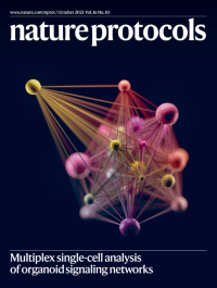
Organoid PTM signaling network from the TOBis MC protocolA post-translational modification (PTM) signaling network of organoid enterocytes derived from TOBis mass cytometry data. The size of each node represents an individual PTM earth mover’s distance (EMD) score, and the edges represent the density-resampled estimation of mutual information (DREMI) score connecting each node.
See Sufi et al.
-
No. 9 September 2021
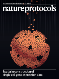
Cells shaped like tiles fall into place to reconstruct their tissue of originnovoSpaRc reconstructs tissues and their associated spatial gene expression patterns based on the information encoded by single cells that were dissociated from such tissues. It is similar to charting a map or completing a puzzle—the cells are analogous to the individual puzzle pieces that together make up the whole picture, or tissue.
See Moriel et al.
-
No. 8 August 2021
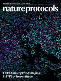
CODEX imaging of human tonsilA formalin-fixed paraffin-embedded tissue section was imaged with the CODEX multiplexed imaging platform. Select markers (CD3–cyan, CD20–blue, CD31–white, CD56–green, CD68–magenta, Ki-67–red and cytokeratin–yellow) are highlighted.
See Black et al.
-
No. 7 July 2021
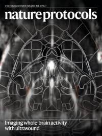
Imaging whole-brain activity with ultrasoundFunctional ultrasound imaging (fUS), a hemodynamic-based method, provides a readout of whole-brain activity in awake mice at high spatiotemporal resolution. The image displays a transverse view of the mouse brain microvasculature, captured with fUS. The fUS volume was registered in the Allen Common Coordinate Framework (outlines) using open-source software facilitating the analysis of ~250 brain regions across subjects.
SeeBrunner et al.
-
No. 6 June 2021
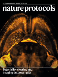
Tutorial for clearing and imaging tissue samplesA CLARITY-cleared Thy-1-YFP-H mouse brain imaged with a mesoSPIM (mesoscale selective plane illumination initiative) light-sheet microscope. The image shows a single plane out of a 3D stack covering the whole brain.
See Weiss et al.
-
No. 5 May 2021
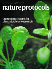
ArabidopsisIn this issue, Kremer et al. describe two peat-based gnotobiotic growth platforms that can be used to support growth of Arabidopsis thaliana in the presence or absence of microorganisms.
See Kremer et al.
-
No. 4 April 2021
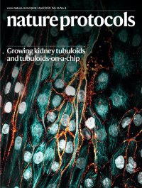
Apical view on a kidney tubuloid cell–derived tubule cultured under perfusion flow in a microfluidic platform (the OrganoPlate).The image displays immunofluorescence staining of the nuclei (white), sodium-potassium-ATPase (orange) and microtubules (cyan).
See Gijzen et al.
-
No. 3 March 2021
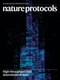
The PAM requirements of hundreds of CRISPR–Cas enzymes profiled with HT-PAMDA depict the expanding capabilities of genome-editing technologies.
See Walton et al.
-
No. 2 February 2021
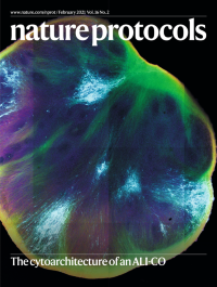
The cytoarchitecture of an ALI-COThe cover shows an air–liquid interface cerebral organoid (ALI-CO) expressing foci of a membrane-targeted GFP construct (cyan) and stained for the pan-neuronal cytoskeletal marker TUBB3 (red), the axonal marker SMI312 (green) and the neuronal transcription factor NeuroD2 (blue). Thick axon bundles project around the edges and toward the center of the organoid, outlining lobules containing neuronal nuclei and axons in a radially ordered lattice.
See Giandomenico et al.
-
No. 1 January 2021
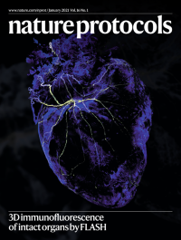
Nervous heart.The cover shows 3D FLASH immunofluorescence staining of nerves (tyrosine hydroxylase) and extracellular matrix (collagen IV) of a whole murine heart.
See Messal et al.