
Volume 16 Issue 11, November 2021
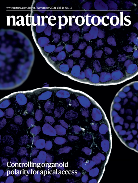
Apical-out human enteroid
Enteroids grown in extracellular matrix (ECM) scaffolds develop apical-in polarity, with the microvilli facing the internal lumen. We reversed enteroid polarity in suspension culture through removal of the ECM. The cover image shows a human colonoid with apical-out polarity; note that the microvilli face the outer surface. Nuclei are stained with DAPI (blue) and the actin cytoskeleton with phalloidin (white).
See Co et al.
Image: Julia Y. Co, Stanford University. Cover design: Tulsi Voralia
Review Articles
-
Advertisement
