Thank you for visiting nature.com. You are using a browser version with limited support for CSS. To obtain
the best experience, we recommend you use a more up to date browser (or turn off compatibility mode in
Internet Explorer). In the meantime, to ensure continued support, we are displaying the site without styles
and JavaScript.
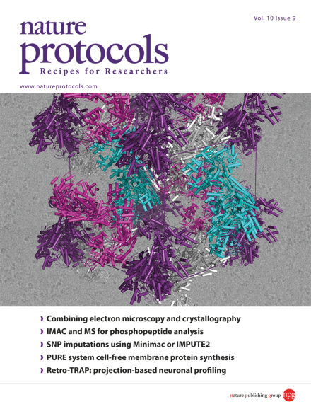
The X-ray crystal structure of Cascade (CRISPR-associated complex for antiviral defense) was determined using a hybrid approach that integrates data from electron microscopy. Cascade assemblies, colored purple, pink, gray, and cyan, are packed into the crystallographic unit cell, and are displayed over the top of an electron micrograph. Based on the protocol by Ryan N. Jackson et al. DOI: 10.1038/nprot.2015.069. Cover design by Jamel Wooten.
Molecular replacement is often used to solve the “phase problem” in X-ray crystallography. This protocol explains how low-resolution maps generated using electron microscopy can be used for the molecular replacement step in solving X-ray structures.
This protocol describes how to perform SNP imputations for GWAS meta-analysis with the Genome of the Netherlands reference panel using Minimac or IMPUTE2.
This protocol describes a technique for measuring protein accumulation on chromatin that combines classical immunofluorescence microscopy detection with flow cytometry.
Determining the phosphorylation of low-abundance peptides in small amount of clinical samples is challenging. In this approach, in-house construction of IMAC columns plus esterification of the peptides are the key features that improve sensitivity.
Nectow, Ekstrand and Friedman describe Retro-TRAP, an extension of the bacTRAP method, for projection-specific ribosome profiling of neurons. This two-component GFP-Nanobody system can, in theory, be adapted to analyze any GFP-expressing cell.
This protocol enables cell-free synthesis of membrane proteins using the PURE system, subsequent quantification of products and analyses of membrane localization efficiency, product orientation, and complex formation in the membrane.
This protocol describes how to generate large quantities of cardiomyocytes from human pluripotent stem cells using a bioreactor. Cells are differentiated by application of Wnt pathway modulators while growing in suspension culture.
Silk-collagen protein scaffolds are seeded with rat primary cortical neurons, which grow neuronal projections that form neuronal networks and functional connectivity.
Sumoylation is a post-translational modification involved in regulating many cellular processes. This protocol is for detecting sumoylation sites in cultured cells using a diGly-Lys (K-ɛ-GG)–specific antibody and mass spectrometry–based proteomics.
This protocol describes flow cytometry panels that can be used to analyze and isolate dendritic cell (DC) progenitors including granulocyte, monocyte and DC progenitor cells; monocyte and DC progenitor cells; and common DC progenitor and DC precursor cells.
18F is commonly used for preparation of probes for position-emission tomography. This protocol is for the modular synthesis of radiolabeling precursors via a copper-catalyzed 'click' reaction followed by a one-step 18F labeling.
EmRiboSeq determines the precise location of embedded ribonucleotides in the S. cerevisiae genome, tracking DNA polymerase activity in vivo. An adaptation of this protocol, EndoSeq, also allows the genome-wide mapping of other noncanonical bases.
Single-cell analysis has shown that a lot of information can be lost by analyzing homogenates of tissues. This protocol describes how to remove the contents of a single plant cell and directly analyze the metabolites by mass spectrometry.