Thank you for visiting nature.com. You are using a browser version with limited support for CSS. To obtain
the best experience, we recommend you use a more up to date browser (or turn off compatibility mode in
Internet Explorer). In the meantime, to ensure continued support, we are displaying the site without styles
and JavaScript.
NanoLuciferase- and HaloTag-based screening technologies are versatile tools suitable for the live-cell analysis of the entire small-molecule-induced degradation cascade to uncover the mode of action of proximity-inducing compounds such as PROTACs.
This protocol describes the preparation of long-lasting aggregation-induced emission-based, near-infrared afterglow luminescence nanoprobes. Their enhanced afterglow intensity results in improved imaging sensitivity and depth in vivo.
It can be challenging to obtain meaningful and accurate structural information for air-sensitive proteins. This protocol describes the application of customized vacuum manifold and anaerobic chamber setups for the purification and cryo-electron microscopy analysis of air-sensitive nitrogenase enzymes.
Attempts to reproduce the computational steps described in published omics research often fail. This review provides guidelines for the packaging and containerization of software so that readers can use the exact programs used in published work.
Our authors are invited to write blog posts that describe how they conceived and developed their protocols, prior to publication at Nature Protocols. These stories are published on a community website for researchers who are interested in techniques and methods.
This Collection highlights articles on how to improve methodological clarity in science publications from a spectrum of different journals and subject areas.
Ancient proteins carry genetic information from fossils that are too old or degraded for ancient DNA recovery. This protocol describes the extraction and tandem mass spectrometry sequencing of million-year-old dental enamel proteins for phylogenetic inference.
This protocol details methods for using methanol in methylation reactions, including the synthesis of suitable transition metal-containing catalysts, and in the synthesis of heterocycles. The methods described produce only H2 and H2O as by-products.
Isotope ratio mass spectrometry as described in this protocol can be used to determine natural variation in the abundance of stable isotopes in individual compounds to provide information relevant to metabolism, ecology or climate change.
Surface photovoltage microscopy as described in this protocol allows high spatial and energy resolution mapping of surface-charge distributions on photocatalyst particles, enabling rational design of improved materials.
NanoLuciferase- and HaloTag-based screening technologies are versatile tools suitable for the live-cell analysis of the entire small-molecule-induced degradation cascade to uncover the mode of action of proximity-inducing compounds such as PROTACs.
This protocol describes the preparation of long-lasting aggregation-induced emission-based, near-infrared afterglow luminescence nanoprobes. Their enhanced afterglow intensity results in improved imaging sensitivity and depth in vivo.
A protocol for the generation of induced blastoids, an in vitro integrated model of the human blastocyst derived via somatic reprogramming. This model overcomes restrictions associated with the use of human blastocysts in embryology research.
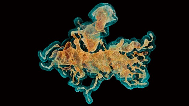
Activated neutrophils labeled with NIR-II lanthanide downshifting nanoparticles can be sequentially imaged through the intact skull of a mouse model of ischemic stroke during adhesion, crawling and extravasation processes
This protocol describes the establishment of a reversible replication barrier using plasmid templates containing a lacO array bound by LacR repressor. The method allows fine control of replication fork movement and replication fork encounter with DNA lesions.