
Optical imaging for diagnostics
The evolution of optical technologies in the context of diagnostic medical imaging has revolutionized, over the past two decades, the way we understand, detect and treat disease. By using labelled tags (which can be tailored to carry a wide range of molecular motifs) and advanced technologies that enable the detection of probe emissions with high sensitivity and specificity, bench researchers and clinicians have been able to diagnose disease by detecting, for example, cancer biomarkers, cell metabolic state and atherosclerotic lesions.
Mostly owing to ease of use and wide applicability range, optical imaging technologies are currently able to provide molecular-grade information, even in operating theatres. These features align well with the drive for personalized health care, envisaged for diagnosing patient subpopulations with increased precision.
This Collection brings together recent efforts in optical diagnostic imaging, and highlights the path bringing the development of optical imaging technologies towards their eventual impact in the clinic. The curated set of research Articles, Reviews, Perspectives and Comments bridge molecular imaging, protein engineering, nanoparticle design and materials science to deliver optical-imaging applications in clinical diagnostics.
The material — of which selected content is free to access until February 10th 2017 — has been published within the last two years in Nature Biomedical Engineering, Nature Biotechnology, Nature Chemical Biology, Nature Communications, Nature Materials, Nature Medicine, Nature Methods, Nature Photonics, Nature Protocols, Nature Reviews Cardiology, Nature Reviews Gastroenterology & Hepatology and Nature Reviews Urology.
Design of banner and header-image: Laura Marshall / Nature Research

In the Photonics Lab
The impact of optical imaging in biomolecular sciences and medical diagnostics is deeply rooted in our understanding of classical electromagnetism and optics. Our fascination with light goes back centuries, even as early as the Neolithic era, when solar emissions were used to measure (time) and predict (seasonal) events. Isaac Newton first described how white light refracts when it crosses a new medium, where the incident beam angle and the refractive index of the media define the qualitative properties with which light is effectively bent. Thomas Young, a physician and polymath, was the first to experimentally prove that light propagates as a wave. Young’s wave theory of light is the pillar for the subsequent works of Hermann von Helmholtz in optics and of James Clerk Maxwell, whose partial differential equations approximate light-matter interactions and derive the absolute speed of light (which in turn is a cornerstone parameter of general relativity).
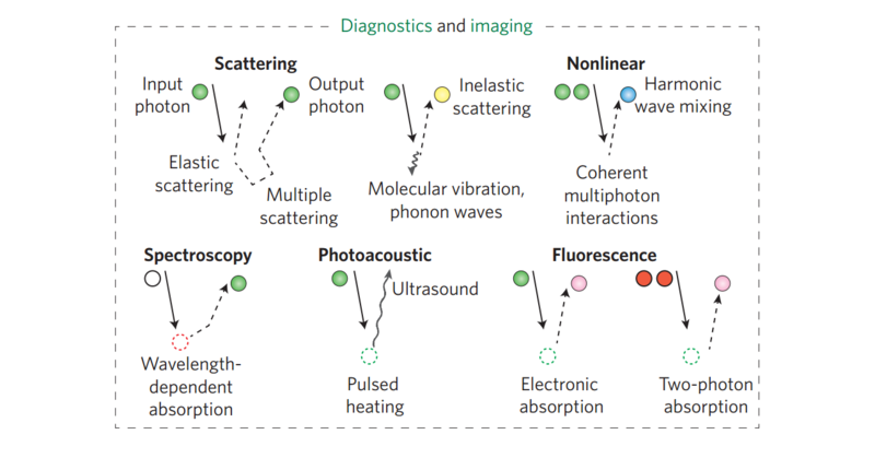
Today’s optical diagnostic systems all adopt these principles. Ultimately, optical diagnostic imaging is based on light: (i) entering a tissue, using a source such as a laser, (ii) interacting with it, typically by diffraction, refraction, scattering and/or absorption, and (iii) being collected via optics or electro-optical sensors.
Harnessing light in living tissue, such as neural circuitry, therefore relies on controlling at least some of the dynamical properties of the electromagnetic waves. Recent research has focussed, for example, on understanding the spin-Hall effect and the spin-orbit interactions of light, as well as toroidal dipole electromagnetic excitations in metamaterials.
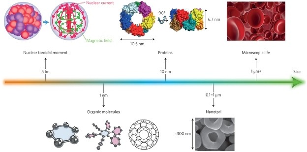
These metamaterials are of particular interest in imaging, not least for their potential to engineer new tools, such as that of a miniaturised camera, measuring just 1.6 mm × 1.6 mm × 1.7 mm, and capable of producing wide-angle images at near-diffraction-limited resolution. The ability to modify the optical field, known as wavefront-shaping, before it enters a scattering medium has further enabled the generation of micrometre-scale focal spots inside tissue.
Such technologies push the limits of optical imaging towards single-molecule detection, where both labelled (such as fluorescent) and label-free (such as Raman spectroscopy) techniques can be coupled to microscopes. The electron microscope has also benefited from the combined use of coherent light sources and fluorescence emissions for the detection of molecules at the nanometre scale. The observation of dynamic events of single molecules at such resolutions can be achieved using super-resolution microscopy, a technique that is playing a major role in discovering, for example, how our genome is capable of organizing itself in architecturally active chromatin structures or how protein chaperone nanomachines are assembled.
