


 Table 1
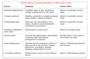


« Prev Next »
Because DNA is the repository of genetic information in each living cell, its integrity and stability are essential to life. DNA, however, is not inert; rather, it is a chemical entity subject to assault from the environment, and any resulting damage, if not repaired, will lead to mutation and possibly disease. Perhaps the best-known example of the link between environmental-induced DNA damage and disease is that of skin cancer, which can be caused by excessive exposure to UV radiation in the form of sunlight (and, to a lesser degree, tanning beds). Another example is the damage caused by tobacco smoke, which can lead to mutations in lung cells and subsequent cancer of the lung. Beyond environmental agents, DNA is also subject to oxidative damage from byproducts of metabolism, such as free radicals. In fact, it has been estimated that an individual cell can suffer up to one million DNA changes per day (Lodish et al., 2005).
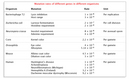
In addition to genetic insults caused by the environment, the very process of DNA replication during cell division is prone to error. The rate at which DNA polymerase adds incorrect nucleotides during DNA replication is a major factor in determining the spontaneous mutation rate in an organism. While a "proofreading" enzyme normally recognizes and corrects many of these errors, some mutations survive this process. Estimates of the frequency at which human DNA undergoes lasting, uncorrected errors range from 1 x 10⁻⁴ to 1 x 10⁻⁶ mutations per gamete for a given gene. A rate of 1 x 10⁻⁶ means that a scientist would expect to find one mutation at a specific locus per one million gametes. Mutation rates in other organisms are often much lower (Table 1).
One way scientists are able to estimate mutation rates is by considering the rate of new dominant mutations found at different loci. For example, by examining the number of individuals in a given population who were diagnosed with neurofibromatosis (NF1, a disease caused by a spontaneous—or noninherited—dominant mutation), scientists determined that the spontaneous mutation rate of the gene responsible for this disease averaged 1 x 10⁻⁴ mutations per gamete (Crowe et al., 1956). Other researchers have found that the mutation rates of other genes, like that for Huntington's disease, are significantly lower than the rate for NF1. The fact that investigators have reported different mutation rates for different genes suggests that certain loci are more prone to damage or error than others.
DNA Repair Mechanisms and Human Disease
DNA repair processes exist in both prokaryotic and eukaryotic organisms, and many of the proteins involved have been highly conserved throughout evolution. In fact, cells have evolved a number of mechanisms to detect and repair the various types of damage that can occur to DNA, no matter whether this damage is caused by the environment or by errors in replication. Because DNA is a molecule that plays an active and critical role in cell division, control of DNA repair is closely tied to regulation of the cell cycle. (Recall that cells transit through a cycle involving the G1, S, G2, and M phases, with DNA replication occurring in the S phase and mitosis in the M phase.) During the cell cycle, checkpoint mechanisms ensure that a cell's DNA is intact before permitting DNA replication and cell division to occur. Failures in these checkpoints can lead to an accumulation of damage, which in turn leads to mutations.
Defects in DNA repair underlie a number of human genetic diseases that affect a wide variety of body systems but share a constellation of common traits, most notably a predisposition to cancer (Table 2). These disorders include ataxia-telangiectasia (AT), a degenerative motor condition caused by failure to repair oxidative damage in the cerebellum, and xeroderma pigmentosum (XP), a condition characterized by sensitivity to sunlight and linked to a defect in an important ultraviolet (UV) damage repair pathway. In addition, a number of genes that have been implicated in cancer, such as the RAD group, have also been determined to encode proteins critical for DNA damage repair.
UV Damage, Nucleotide Excision Repair, and Photoreactivation
As previously mentioned, one important DNA damage response (DDR) is triggered by exposure to UV light. Of the three categories of solar UV radiation, only UV-A and UV-B are able to penetrate Earth's atmosphere. Thus, these two types of UV radiation are of greatest concern to humans, especially as continuing depletion of the ozone layer causes higher levels of this radiation to reach the planet's surface.
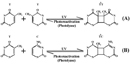
UV radiation causes two classes of DNA lesions: cyclobutane pyrimidine dimers (CPDs, Figure 1) and 6-4 photoproducts (6-4 PPs, Figure 2). Both of these lesions distort DNA's structure, introducing bends or kinks and thereby impeding transcription and replication. Relatively flexible areas of the DNA double helix are most susceptible to damage. In fact, one "hot spot" for UV-induced damage is found within a commonly mutated oncogene, the p53 gene.
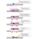
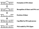
CPDs and 6-4 PPs are both repaired through a process known as nucleotide excision repair (NER). In eukaryotes, this complex process relies on the products of approximately 30 genes. Defects in some of these genes have been shown to cause the human disease XP, as well as other conditions that share a risk of skin cancer that is elevated about a thousandfold over normal. More specifically, eukaryotic NER is carried out by at least 18 protein complexes via four discrete steps (Figure 3): detection of damage; excision of the section of DNA that includes and surrounds the error; filling in of the resulting gap by DNA polymerase; and sealing of the nick between the newly synthesized and older DNA (Figure 4). In bacteria (which are prokaryotes), however, the process of NER is completed by only three proteins, named UvrA, UvrB, and UvrC.
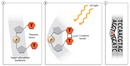
Bacteria and several other organisms also possess another mechanism to repair UV damage called photoreactivation. This method is often referred to as "light repair," because it is dependent on the presence of light energy. (In comparison, NER and most other repair mechanisms are frequently referred to as "dark repair," as they do not require light as an energy source.) During photoreactivation, an enzyme called photolyase binds pyrimidine dimer lesions; in addition, a second molecule known as chromophore converts light energy into the chemical energy required to directly revert the affected area of DNA to its undamaged form. Photolyases are found in numerous organisms, including fungi, plants, invertebrates such as fruit flies, and vertebrates including frogs. They do not appear to exist in humans, however (Sinha & Hader, 2002).
Additional DNA Repair mechanisms
NER and photoreactivation are not the only methods of DNA repair. For instance, base excision repair (BER) is the predominant mechanism that handles the spontaneous DNA damage caused by free radicals and other reactive species generated by metabolism. Bases can become oxidized, alkylated, or hydrolyzed through interactions with these agents. For example, methyl (CH3) chemical groups are frequently added to guanine to form 7-methylguanine; alternatively, purine groups may be lost. All such changes result in abnormal bases that must be removed and replaced. Thus, enzymes known as DNA glycosylases remove damaged bases by literally cutting them out of the DNA strand through cleavage of the covalent bonds between the bases and the sugar-phosphate backbone. The resulting gap is then filled by a specialized repair polymerase and sealed by ligase. Many such enzymes are found in cells, and each is specific to certain types of base alterations.
Yet another form of DNA damage is double-strand breaks, which are caused by ionizing radiation, including gamma rays and X-rays. These breaks are highly deleterious. In addition to interfering with transcription or replication, they can lead to chromosomal rearrangements, in which pieces of one chromosome become attached to another chromosome. Genes are disrupted in this process, leading to hybrid proteins or inappropriate activation of genes. A number of cancers are associated with such rearrangements. Double-strand breaks are repaired through one of two mechanisms: nonhomologous end joining (NHEJ) or homologous recombination repair (HRR). In NHEJ, an enzyme called DNA ligase IV uses overhanging pieces of DNA adjacent to the break to join and fill in the ends. Additional errors can be introduced during this process, which is the case if a cell has not completely replicated its DNA in preparation for division. In contrast, during HRR, the homologous chromosome itself is used as a template for repair.
Mutations in an organism's DNA are a part of life. Our genetic code is exposed to a variety of insults that threaten its integrity. But, a rigorous system of checks and balances is in place through the DNA repair machinery. The errors that slip through the cracks may sometimes be associated with disease, but they are also a source of variation that is acted upon by longer-term processes, such as evolution and natural selection.
References and Recommended Reading
Branze, D., & Foiani, M. Regulation of DNA repair throughout the cell cycle.
Nature Reviews Molecular Cell Biology 9, 297–308 (2008) doi:10.1038/nrm2351.pdf (link to article)
Crowe, F. W., et al. A Clinical, Pathological, and Genetic Study of Multiple Neurofibromatosis (Springfield, Illinois, Charles C. Thomas, 1956)
Lodish, H., et al. Molecular Biology of the Cell, 5th ed. (New York, Freeman, 2004)
Sinha, R. P., & Häder, D. P. UV-induced DNA damage and repair: A review. Photochemical and Photobiological Sciences 1, 225–236 (2002)


