Thank you for visiting nature.com. You are using a browser version with limited support for CSS. To obtain
the best experience, we recommend you use a more up to date browser (or turn off compatibility mode in
Internet Explorer). In the meantime, to ensure continued support, we are displaying the site without styles
and JavaScript.
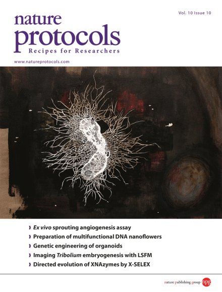
The cover image is reproduced from a mixed oil and acrylic on canvas painting by Jestin George. The image depicts the artist's interpretation of the immunostained microvascular network generated during the metatarsal angiogenesis assay. Jestin George is a research technician working on the LRG1 project in the Department of Cell Biology, University College London Institute of Ophthalmology, London, UK. Based on the Protocol by Weihua Song et al. doi: 10.1038/nprot.2015.097.
This protocol describes how to set up an assay investigating vessel outgrowth from mouse fetal metatarsals. This assay is an ex vivo assay to investigate sprouting angiogenesis.
This protocol describes how to grow untransformed human colonic organoids and deliver genes of interest into the organoids via the piggyBac transposon or gene editing using the CRISPR-Cas9 system.
The Stelzer lab describes how to live-image Tribolium embryos using light-sheet-based fluorescence microscopy (LSFM). Imaging can proceed for up to 120 h, allowing the entire embryonic development of this important model insect to be recorded.
This protocol describes how to prepare multifunctional DNA nanoflowers. Rolling-circle amplification of a designed template containing drug binding, cell targeting, and fluorescent dye binding sites creates structures of controllable size for drug delivery.
The Gonzalez laboratory provides its allograft protocol for studying tumorigenesis in Drosophila. Transplanting tissue from donor larvae to adult hosts allows its tumorigenic potential to be determined.
The study of meiosis in plants is considered to be gender-biased owing to the easy accessibility of male meiocytes. This protocol describes how to prepare and image female Arabidopsis meiocytes to investigate protein localization during meiosis.
In this protocol, neuronal miRNAs and transcription factors are used to directly convert human fibroblasts to striatal medium spiny neurons, a neuronal subtype important in motor control and the main cell type affected in Huntington's disease.
Unbiased proteome-level discovery of intracellular drug targets can be achieved by plotting melting curves of proteins from untreated and drug-treated cells. Multiplexed quantitative mass spectrometry using TMT10 reagents makes this possible.
This protocol uses CuAAC click chemistry along with two different methods for expansion of the genetic code to assemble protein-protein conjugates for studying the post-translational modification of proteins by ubiquitin (ubiquitylation).
In this protocol, limb muscles are physically and enzymatically dissociated to maximally release resident mononucleated cells. Pure populations of either quiescent or activated muscle stem cells are then isolated by flow cytometry.
The authors describe methods for the directed evolution of artificial endonuclease and ligase enzymes by X-SELEX, from diverse repertoires of synthetic genetic polymers (XNAzymes). The protocol has been applied to four different XNA chemistries and three different reactions, and it is, in principle, applicable to many more.