Thank you for visiting nature.com. You are using a browser version with limited support for CSS. To obtain
the best experience, we recommend you use a more up to date browser (or turn off compatibility mode in
Internet Explorer). In the meantime, to ensure continued support, we are displaying the site without styles
and JavaScript.
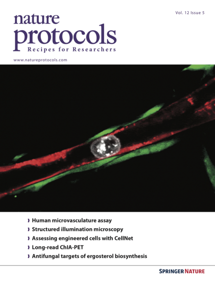
An MDA-MB-231 tumor cell (white) arrested in a capillary (red) sheathed with pericytes (green), visualized within an on-chip human microvascular network. Image taken from the protocol by Kamm et al. doi:10.1038/nprot.2017.018. Cover design by Jamel Wooten.
This protocol describes how to set up and use an in vitro model of the human microcirculation with the capability to recapitulate discrete steps of early metastatic seeding, including tumor cell arrest and transendothelial migration.
This protocol enables the study of protein complexes by trapping them in virus-like particles and thereby preserving cellular integrity. The Virotrap protocol is complemented by SFINX, an online data analysis tool for interactomics studies.
Li et al. provide a protocol for long-read ChIA-PET, a technique for mapping chromatin interactions. The longer paired-end tags, which are generated by tagmentation, provide sufficient coverage to determine haplotype-specific chromatin interactions at single-nucleotide resolution.
Four different techniques for preparing and acquiring super-resolution CLEM data sets on aldehyde-fixed specimens are provided: Tokuyasu cryosectioning; whole-cell mount; cell unroofing and platinum replication; and resin embedding and sectioning.
By combining minimal inhibitory concentration testing with GC–MS-based pathway analysis, it is possible to identify which enzymes in the ergosterol biosynthesis pathway are being targeted by potential new antifungal substances.
This protocol describes how to produce molecularly imprinted polymers, synthetic receptors that can be produced with antibody-like binding properties. They are easier and cheaper to produce than antibodies and lectins.
This protocol describes the preparation of calibration bead slides, their use and additional strategies to reduce artifacts of structured illumination microscopy that will allow researchers to exploit the technique's full potential for biological applications.
This protocol describes how to prepare samples for labeling nuclei of cultured mammalian cells for 3D structured illumination microscopy of nuclear structures. Image acquisition, registration and downstream image analysis are also described.
Expandable and multipotent induced cardiac progenitor cells (iCPCs) are generated from mouse adult fibroblasts using forced expression of Mesp1, Tbx5, Gata4, Nkx2.5 and Baf60c and activation of the Wnt and JAK/STAT signaling pathway. Furthermore, embryonic potency of iCPCs is tested ex vivo.
Bioluminescence and radioluminescence are both dim phenomena that require a highly sensitive microscope for imaging at the cellular level. This protocol describes how to construct a modular low-light microscope for imaging these events.
This protocol describes how to generate a human 3D lymphatic capillary network from primary cells without the use of synthetic scaffolds or exogenous factors. The tissue is stable over many weeks and accurately recapitulates in vivo human dermal lymphatic microvasculature.
CellNet is a computational platform designed to improve cell fate engineering protocols. Here the authors demonstrate how to apply CellNet to RNA-seq data and how to build a completely new CellNet platform applicable to new species or cell types/tissues.