Thank you for visiting nature.com. You are using a browser version with limited support for CSS. To obtain
the best experience, we recommend you use a more up to date browser (or turn off compatibility mode in
Internet Explorer). In the meantime, to ensure continued support, we are displaying the site without styles
and JavaScript.
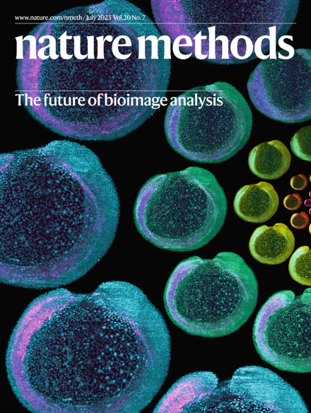
Advanced bioimage analysis tools are poised to disrupt the way in which microscopy images are acquired and analyzed. This Focus issue shares the hopes and opinions of experts on the near and distant future of image analysis.
Astyanax mexicanus exists in two forms: a surface form that is abundantly distributed throughout freshwater bodies in Middle America and a blind subterranean form endemic to caves in northeastern Mexico. These diverse fish populations have become a vertebrate model for investigating the genetic basis of environmental adaptation.
The field of bioimage analysis is poised for a major transformation, owing to advancements in imaging technologies and artificial intelligence. The emergence of multimodal foundation models — which are akin to large language models (such as ChatGPT) but are capable of comprehending and processing biological images — holds great potential for ushering in a revolutionary era in bioimage analysis.
In the ever-evolving landscape of biological imaging technology, it is crucial to develop foundation models capable of adapting to various imaging modalities and tackling complex segmentation tasks.
Concurrent advances in imaging technologies and deep learning have transformed the nature and scale of data that can now be collected with imaging. Here we discuss the progress that has been made and outline potential research directions at the intersection of deep learning and imaging-based measurements of living systems.
Advanced imaging techniques provide holistic observations of complicated biological phenomena across multiple scales while posing great challenges to data analysis. We summarize recent advances and trends in bioimage analysis, discuss current challenges toward better applicability, and envisage new possibilities.
We dream of a future where light microscopes have new capabilities: language-guided image acquisition, automatic image analysis based on extensive prior training from biologist experts, and language-guided image analysis for custom analyses. Most capabilities have reached the proof-of-principle stage, but implementation would be accelerated by efforts to gather appropriate training sets and make user-friendly interfaces.
I share my opinions on the benefits of and bottlenecks for hyperspectral and time-resolved imaging. I also discuss current and future perspectives for analyzing these types of data using the phasor approach.
A key step toward biologically interpretable analysis of microscopy image-based assays is rigorous quantitative validation with metrics appropriate for the particular application in use. Here we describe this challenge for both classical and modern deep learning-based image analysis approaches and discuss possible solutions for automating and streamlining the validation process in the next five to ten years.
The bridging of domains such as deep learning-driven image analysis and biology brings exciting promises of previously impossible discoveries as well as perils of misinterpretation and misapplication. We encourage continual communication between method developers and application scientists that emphases likely pitfalls and provides validation tools in conjunction with new techniques.
The future of bioimage analysis is increasingly defined by the development and use of tools that rely on deep learning and artificial intelligence (AI). For this trend to continue in a way most useful for stimulating scientific progress, it will require our multidisciplinary community to work together, establish FAIR (findable, accessible, interoperable and reusable) data sharing and deliver usable and reproducible analytical tools.
The language used by microscopists who wish to find and measure objects in an image often differs in critical ways from that used by computer scientists who create tools to help them do this, making communication hard across disciplines. This work proposes a set of standardized questions that can guide analyses and shows how it can improve the future of bioimage analysis as a whole by making image analysis workflows and tools more FAIR (findable, accessible, interoperable and reusable).
Here we discuss the prospects of bioimage analysis in the context of the African research landscape as well as challenges faced in the development of bioimage analysis in countries on the continent. We also speculate about potential approaches and areas of focus to overcome these challenges and thus build the communities, infrastructure and initiatives that are required to grow image analysis in African research.
Wrangling big data is now part of being a biomedical scientist, and mandates on data sharing have entered the scene. Mandates can alter behavior, but data sharing also needs incentives and shifts in science culture.
Three groundbreaking studies have created a new generation of genetically encoded voltage indicators, empowering us to tackle a host of questions on our path toward understanding the brain.
Genome architecture mapping (GAM) enables understanding of 3D genome structure in the nucleus. We directly compared multiplex-GAM and Hi-C data and found that local chromatin interactions were generally detected by both methods, but active genomic regions rich in enhancers that established higher-order contacts were preferentially detected by GAM.
Our study introduces conditional autoencoder for multiplexed pixel analysis (CAMPA), a deep-learning framework that uses highly multiplexed imaging to identify consistent subcellular landmarks across heterogeneous cell populations and experimental perturbations. Generating interpretable cellular phenotypes revealed links between subcellular organization and perturbations of RNA production, RNA processing and cell size.
Cell type-specific transgene expression in mice has broad utility in biomedical research. We developed a versatile system for in vivo transgene delivery using adeno-associated virus (AAV). Efficient and tissue-specific transgene expression is achieved by regulating the expression of the gene encoding the AAV receptor, thereby precisely targeting AAV to the cell type of interest.
This updated analysis of the Cell Tracking Challenge explores how algorithms for cell segmentation and tracking in both 2D and 3D have advanced in recent years, pointing users to high-performing tools and developers to open challenges.
This paper proposes two new anisotropy metrics—the Fourier shell occupancy and the Bingham test—that can be used to understand the quality of cryogenic electron microscopy maps.
The miniature RNA-guided endonuclease IscB and its ωRNA were engineered for efficient gene editing in mammalian cells. Fusions of ‘enIscB’ to T5 exonuclease and cytosine or adenosine deaminase yield versatile tools for genome engineering.
MISAR-seq combines spatial-ATAC-seq and RNA-seq for spatial profiling of both chromatin accessibility and gene expression, as demonstrated in the developing mouse brain.
CAMPA (Conditional Autoencoder for Multiplexed Pixel Analysis) learns representations of molecular pixel profiles from multiplexed images that can be clustered to quantify subcellular landmarks and capture interpretable cellular phenotypes.
The Photopick platform, which can be used for phenotype-activated cell selection, was used to develop the improved voltage sensors QuasAr6a and QuasAr6b. These GEVIs offer improved signals and are useful for all-optical electrophysiology.
A suite of tools including positive-going voltage indicators, a high-speed two-photon microscope, and denoising software enables prolonged imaging of electrical activity in neurons with limited toxicity.
The ASAP4 family of genetically encoded voltage indicators allows recording of action potentials and subthreshold activity with either one- or two-photon microscopy over extended periods of time.