Thank you for visiting nature.com. You are using a browser version with limited support for CSS. To obtain
the best experience, we recommend you use a more up to date browser (or turn off compatibility mode in
Internet Explorer). In the meantime, to ensure continued support, we are displaying the site without styles
and JavaScript.
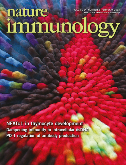
Nuclear translocation of NFAT proteins is necessary for gene expression. Serfling and colleagues (p127; News and Views by Macian, p116) show that in pre-TCR–negative thymocytes, IL-7–Jak3–mediated signals result in nuclear localization of NFA Tc1. The original image by Amiya K. Patra shows nuclear NFA Tc2 (yellow, a merge of red (NFA Tc2) and green (DAPI)) in COS-7 cells transfected with a vector expressing NFA Tc2. Artwork by Lewis Long.
Signaling via the complement factors C3a and C5a regulates effector T cell responses. Evidence now links the absence of local complement activation with a default pathway that leads to the polarization of Foxp3+ regulatory T cells.
Maintenance of the peripheral naive CD8+ T cell pool depends on T cell antigen receptor (TCR) signals and interleukin 7 (IL-7). TCR signaling limits the duration of IL-7 signals to avoid an interferon-γ-mediated mechanism of apoptotic death of naive CD8+ T cells.
Severe intestinal pathology associated with infection with Toxoplasma gondii is a result of the loss of Paneth cells, an unexpected side effect of the secretion of IFN-γ by protective CD4+ T cells in the intestinal mucosa.
Double-negative (DN) thymocytes depend on interleukin 7 (IL-7) for survival. In DN2 and DN3 cells, a new role has now been identified for signaling via the IL-7 receptor and the kinase Jak3 that leads to noncanonical activation of the transcription factor NFATc1 and upregulation of expression of the antiapoptotic protein Bcl-2.
Interleukin 7 (IL-7) signaling is essential during early lymphocyte development. Patra and colleagues identify a distinct IL-7–kinase Jak3–dependent pathway that activates the transcription factor NFATc1 in DN1 thymocytes to promote their survival.
Immune responses can cause immunopathology. Yarovinsky and colleagues show that IFN-γ induced by T cell–intrinsic TLR signaling in CD4+ TH1 cells during Toxoplasma gondii infection causes dysbiosis and loss of Paneth cells.
Why signaling via both the receptor for interleukin 7 (IL-7R) and the T cell antigen receptor (TCR) is required for T cell homeostasis has been unclear. Singer and colleagues show that engagement of the TCR interrupts IL-7R signals to prevent a mechanism of cytokine-induced cell death.
Follicular regulatory T cells control humoral immune responses, but how these cells are in turn controlled has been unclear. Sharpe and colleagues demonstrate that signaling via PD-1 regulates number and function of these cells.
Complement provides costimulatory signals to T cells. Medof and colleagues demonstrate that an absence of complement signaling in naive T cells generates an autoinductive loop to drive induced regulatory T cells.
The intracellular sensor DDX41 is important for generating innate responses to DNA viruses. Liu et al. demonstrate that the ubiquitin ligase TRIM21 degrades and thereby regulates DDX41-dependent responses.
Hacohen and colleagues use an integrative approach that combines quantitative proteomics, genomics and small molecule perturbations to identify new genes involved in DNA sensing and type I interferon production.