
Volume 85 Issue 10, October 2005
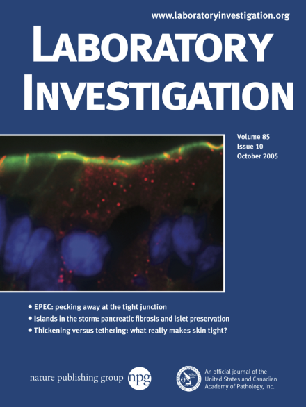
Mouse intestine stained for the tight junction protein occludin (red), f-actin (green), and nuclei (blue; using Hoechst 33342). In contrast to the restricted tight junction localization seen in tissues from control mice, this tissue from enteropathogenic E. coli-infected mouse intestine shows occludin localized to both the tight junction and intracellular vesicles. This occludin redistribution correlated with functional tight junction disruption. See article by Shifflett et al on page 1308 for further details.
Inside Lab Invest
-
Advertisement
