Thank you for visiting nature.com. You are using a browser version with limited support for CSS. To obtain
the best experience, we recommend you use a more up to date browser (or turn off compatibility mode in
Internet Explorer). In the meantime, to ensure continued support, we are displaying the site without styles
and JavaScript.
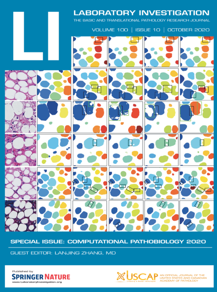
The paper by Roy et al. ( p 1374) shows the use of deep learning for histological assessment of liver biopsies, resulting in accurate quantification of hepatic steatosis. The cover shows visualization of segmented steatosis droplets in masks of distinct colors.
Polarization-second harmonic microscopy was utilized to investigate whether collagen ultrastructure in thyroid due to four carcinoma types and Graves’ disease could be differentiated in human histopathology samples. Three parameters were extracted, revealing that the degree of linear polarization and χ(2)zzz/χ(2)zxx were effective in differentiating some diseases, while the parameter χ(2)xyz/χ(2)zxx was less effective.
Proteomic profiling may contribute to the analysis and classification of cancer. The authors applied the digital western blot technique DigiWest with a panel of 102 proteins and phosphoproteins in combination with a machine learning algorithm to classify the tissue origin of five common cancer types in fresh frozen and formalin-fixed paraffin-embedded tissue. DigiWest profiling represents a valuable method for cancer classification, yielding conclusive and decisive data, thus making this approach attractive for routine clinical applications.
This study aims to classify histopathological images of malignant lymphoma through deep learning. The classifier achieved the high levels of accuracy in comparison to diffuse large B-cell lymphoma, follicular lymphoma, and reactive lymphoid hyperplasia, which were higher than those of pathologists. Artificial intelligence can potentially support diagnosis of malignant lymphoma.
Digital spatial profiling is a new high-plex technology with potential to multiplex hundreds of proteins on a single slide. Here the authors validate the digital aspect of the technology on a control tissue microarray with known amounts of PD-L1 expression to show it has quantitative capacity comparable to quantitative immunofluorescence.
Traditional RNA sequencing data may fail to detect the exact cellular changes in tumor cells. With comprehensively single-cell and traditional RNA-seq data, these authors detected and compared differentially expressed genes in lung adenocarcinoma. These results may improve our understanding of cellular and molecular differences between cancerous and non-malignant tissue and provide tumor markers as well as potential therapeutic targets.
The authors developed a novel simplified assay for glioblastoma transcriptional classification on formalin-fixed-paraffin-embedded tissue samples. On such dataset, immunohistochemical profiles, based on expression of a restricted panel of gene classifiers, were integrated by machine learning approach to generate a glioblastoma transcriptional signature based on protein quantification that allowed to efficiently assign transcriptional subgroups to an extended cohort. Correlations with both histopathological features and clinical outcome have been also performed.
RNA of sufficient quality and quantity can be extracted from formalin-fixed paraffin-embedded (FFPE) samples to obtain comprehensive transcriptome profiling using the 3′ massive analysis of c-DNA ends (MACE) RNA-sequencing technology. Thus, MACE provides an opportunity for utilizing FFPE samples stored in histological archives.
We propose a statistical framework named WTSPP to generate prognostic gene signatures. Using ovarian cancer and lung adenocarcinoma as examples, they provide evidence that our prognostic signatures overperform previous reported signatures, capture prognostic features not explained by clinical variables and expose biologically relevant prognostic pathways.
Accurate quantification of steatosis in liver biopsies is a key step in the treatment of patients with fatty liver diseases. To assist pathologists for such analysis tasks, we develop a novel deep learning-based framework to segment overlapped steatosis droplets in whole slide liver biopsy images. Quantitative measurements of steatosis at both pixel and object-level present strong correlation with clinical data, suggesting its potential for clinical decision support.