Thank you for visiting nature.com. You are using a browser version with limited support for CSS. To obtain
the best experience, we recommend you use a more up to date browser (or turn off compatibility mode in
Internet Explorer). In the meantime, to ensure continued support, we are displaying the site without styles
and JavaScript.
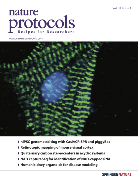
Genome-edited hiPSC differentiated into the cardiomyocyte lineage. An hiPSC line underwent genome editing using a high-efficiency protocol based on a dox-inducible, Cas9-expressing piggyBac transgene. After piggyBac excision, the hiPSC line was differentiated into cardiomyocytes. Green, sarcomeric α-actinin immunostaining; blue, DAPI-stained nucleus. Image taken from the protocol by Pu et al. doi:10.1038/nprot.2016.152. Cover design by Jamel Wooten.
Standard methods for antibody staining of Drosophila larvae have worked poorly or involved dissection. The Doe laboratory describes a reliable protocol for immunofluorescent antibody staining of intact Drosophila larvae that works well for all larval stages.
Different concentrations of activin A and BMP4 are used to polarize stem cells into mesodermal subtypes that reflect mid-primitive-streak cardiogenic mesoderm and posterior-primitive-streak hemogenic mesoderm.
Zilionis et al. describe a protocol for the inDrops platform, which is a high-throughput droplet microfluidics system that allows for single-cell barcoding of over 15,000 cells in 1 h.
This protocol describes the diastereoselective approach for the synthesis of complex, acyclic molecular architectures possessing two stereogenic centers in a 1,4 relationship based on a zirconium-promenade reaction using the Negishi reagent.
This protocol uses a doxycycline-inducible Cas9 transgene carried on a piggyBac transposon for robust, highly efficient and scarless genome editing of human iPSCs, enabling a parental line to be engineered to harbor many separate mutations.
This protocol describes surgical techniques for long-term intrathecal drug administration to rodent CNS tissue and cerebrospinal fluid collection for monitoring of drug concentration and distribution.
Correlative light and electron microscopy (CLEM) promises new insight into cellular processes by combining spatiotemporal information with high-resolution structural information. This protocol describes cryo-CLEM of virus-infected mammalian cells.
Correia-Melo et al. describe a protocol to generate and maintain mitochondria-depleted mammalian cell lines. These cells can be used to investigate the role of mitochondria in various cellular processes such as cell death and senescence.
This protocol describes how to differentiate human pluripotent stem cells into nephron progenitor cells with subsequent generation of 2D and 3D kidney organoids.