
Volume 16 Issue 1, January 2021
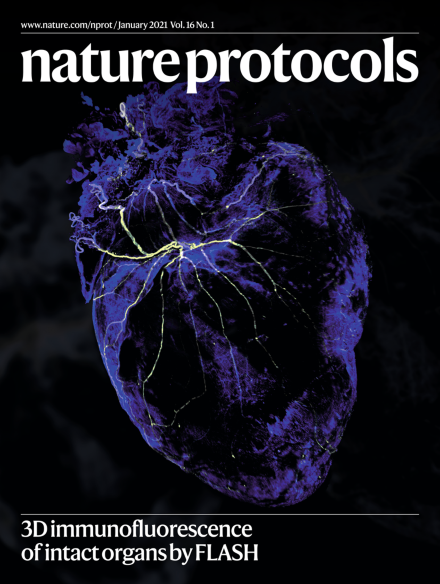
Nervous heart.
The cover shows 3D FLASH immunofluorescence staining of nerves (tyrosine hydroxylase) and extracellular matrix (collagen IV) of a whole murine heart.
See Messal et al.
Image: Axel Behrens. Cover design: Tulsi Voralia.
Review Articles
-
Advertisement
