-
January 18, 2014
Gallery: The Forgotten Asiatic Lion (And Why Po... -
July 04, 2013
Conservation’s Dark Side: How the Bushmen... -
June 18, 2013
Researchers Use Laser Technology and Find a Lon... -
June 16, 2013
What Happens When Artists Look at the Brain? -
June 13, 2013
A Sunken Egyptian City is Rediscovered, Stunnin... -
June 06, 2013
Why Should We Eat Insects? It’s the Futur... -
May 16, 2013
High School "Citizen Scientists'" Work May Help... -
May 06, 2013
CozmicZoom: An App That Will Humble You As You ... -
April 23, 2013
My Grandfather Is In A Sugar-Apple -
April 19, 2013
The Only Positive Effect Of The Cuban Embargo? ... -
March 22, 2013
The Unhappiest Place On Earth (And How Happines... -
March 14, 2013
Tackling Mental Illness In Africa -
March 05, 2013
The Violent History Of Mauritia: Birth, Oblivio... -
February 19, 2013
Is Global Warming Chiefly Manmade? One Graphic ... -
February 14, 2013
Lovers' Hearts Beat At The Same Rate Everyday -
January 22, 2013
My Med Student Friends Are Zombies: On The Comp... -
January 16, 2013
A Story Of Happy And Sad Endings: On An Afflict... -
October 16, 2012
Fukushima Dogs Had Symptoms Comparable To Post-... -
October 03, 2012
Statistics Is The Sexy In Science -
September 18, 2012
New Smart Light Bulb LIFX Might Just Revolution... -
September 14, 2012
Watch New Footage Of Curiosity Landing On Mars ... -
September 11, 2012
Forget Aeolians, We Need Airborne Wind Farms To... -
August 30, 2012
The Internet Is Not Free: We're In The Filter B... -
August 24, 2012
When A Science Writer Turns His Back On Science -
August 16, 2012
When Peer Review Turns Frustrated Authors Into ... -
August 01, 2012
Chronicling The Coming Of Age Of Two Turtledove... -
July 21, 2012
Soccer's Big Data Revolution -
July 11, 2012
Leap Motion: Death Of The Mouse -
July 04, 2012
Higgs Boson: the jokes edition -
June 29, 2012
A Call To Arms For Young Science Journalists -
June 22, 2012
Your Musical Brain -
June 15, 2012
Science With Religion Can Save More Lives -
June 07, 2012
Keep Cool Science Journalists -
May 24, 2012
Son's Cells Linked To Mother's Cancer -
May 10, 2012
Bat-Robot: From Batman To Reality -
May 08, 2012
That The East Is Rising Is Great (Not Bad) News -
May 03, 2012
The Disorder That Killed One Soccer Player And ... -
April 20, 2012
A Rose By Any Other Name Would Look As Red -
April 13, 2012
My Attempt To Further Promote Young Science Wri... -
March 28, 2012
FuturICT: The Tool That Promises To Predict The... -
March 14, 2012
6 Android Apps For Geeks -
March 02, 2012
Lenses On Biology: Scientists And Science Stude... -
February 24, 2012
A Mauritian At Science Online 2012 -
January 03, 2012
Undressing The Brain With Scale -
December 11, 2011
Love You Melbourne -
November 23, 2011
Funny Things Peer-Reviewers Are Saying Behind Y... -
November 05, 2011
What Is The Place Of New Science Bloggers In To... -
October 27, 2011
Scientists Should Remember That Science Is Much... -
October 22, 2011
Of Kindle, Instapaper and Thesis-ing -
September 04, 2011
Encephalon #90 -
August 07, 2011
Define Science -
July 27, 2011
Your Baby On Crack -
July 17, 2011
Are fMRI Telling The Truth? Role Of Astrocytes ... -
July 08, 2011
15 Mistakes Young Researchers Make -
July 05, 2011
Scientific American's New Science Blogging Netw... -
July 03, 2011
Is Google+ The Google Portal? -
July 02, 2011
Social Media: Getting Content That Interests You -
June 02, 2011
In Science, The More The Merrier -
May 15, 2011
Science Blogs Are Good For You -
May 11, 2011
Medical Research In Australia Is Safe -
May 06, 2011
When The Microscope Goes Digital -
May 05, 2011
In Which I Won A Science Blogging Contest -
April 29, 2011
Science On Royal Wedding Day -
April 22, 2011
The Neuroscience Of Smurfs -
April 14, 2011
Medical Research Funding Cutbacks: Proof That S... -
April 10, 2011
Take A Stance Against Medical Research Cutbacks... -
April 03, 2011
Should Extremely Preterm Babies Be Saved? -
March 30, 2011
Dopamine: The Link Between Neuronal Activity An... -
March 21, 2011
Honors Students Are Newbies -
March 18, 2011
Common Ancestry: We Come From One -
February 25, 2011
Google Teaches Me How To Cook -
February 14, 2011
First Time At The Research Department -
January 31, 2011
Brains Breathe: Dopamine's Role In Preterm Infants -
January 27, 2011
More! More! More! -
January 20, 2011
Building Models: When Science Can Be Fun -
January 14, 2011
An Enjoyable Medical Exam -
January 05, 2011
Application Hassles -
December 29, 2010
The Exam Effect -
December 23, 2010
Welcome, welcome! -
December 20, 2010
In which I help out a researcher...
« Prev « Prev Next » Next »


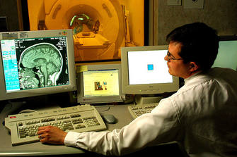
 The prominent rise of late of interdisciplinary fields like cognitive psychology, cognitive neuroscience and neuropsychology is due in no small part to the emergence of the
  In the brain, energy consumption has long been attributed to neural activity. As such, areas fMRI signals have been assumed to represent neural activity. Neural activity produces increases in CBF which occur in seconds and are highly restricted to areas of brain activation. What's the link between neurons and increased CBF? A widely accepted hypothesis is that glutamate receptors, activated during synaptic transmission, cause postsynaptic increases in Ca2+. These eventually set off enzymes which produce vasoactive agents (K+, H+, neurotransmitters, adenosine, nitric oxide, etc). These agents act on receptors on vascular smooth muscles (i.e. on blood vessels) and mediate vasodilatation, indirectly increasing CBF and provision of oxygen and metabolites.


EEG measures voltages that arise due to electrical impulses through neurons. Readings are thus a measure of neuronal activity. Astrocytic activity does not confound these readings, as far as I can tell.
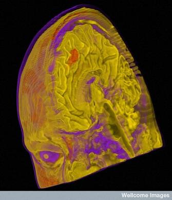
However, EEG is not able to come up with any decent map of neuronal activation. Therein lies the true power of fMRI which is able to show areas of "activation."
What should be done to better interpret fMRI signals is to determine exactly how much of those signals are accounted for my astrocytic and not neuronal activity.
first of all congrats for your very interesting post.
I have just a very basic question. I was wondering if the studies performing EEG and fMRI at the same time could help to solve this "puzzle" related to astrocytes and neuronal activation?
*sorry about my poor english.
Regards.