Volume 11
-
No. 12 December 2014
Cover image supplied by M. J. Gora, V. J. Madden and G. J. Tearney, Wellman Center for Photomedicine at Massachusetts General Hospital and Harvard Medical School, USA. A 3D image of the oesophagus created by rendering data obtained from an unsedated human subject using a swallowable tethered capsule endomicroscopy device. The capsule employs optical coherence tomography: optics within the capsule spin a focused beam around its circumference, acquiring cross-sectional images as it traverses the organ via peristalsis. A flexible tether containing an optical fibre is attached to the capsule and can be used to control its position and to remove it from the mouth so that it can be disinfected and reused.
-
No. 11 November 2014
Cover image supplied by M. J. Gora, V. J. Madden and G. J. Tearney, Wellman Center for Photomedicine at Massachusetts General Hospital and Harvard Medical School, USA. A 3D image of the oesophagus created by rendering data obtained from an unsedated human subject using a swallowable tethered capsule endomicroscopy device. The capsule employs optical coherence tomography: optics within the capsule spin a focused beam around its circumference, acquiring cross-sectional images as it traverses the organ via peristalsis. A flexible tether containing an optical fibre is attached to the capsule and can be used to control its position and to remove it from the mouth so that it can be disinfected and reused.
Focus
-
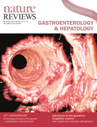
No. 10 October 2014
Cover image supplied by M. J. Gora, V. J. Madden and G. J. Tearney, Wellman Center for Photomedicine at Massachusetts General Hospital and Harvard Medical School, USA. A 3D image of the oesophagus created by rendering data obtained from an unsedated human subject using a swallowable tethered capsule endomicroscopy device. The capsule employs optical coherence tomography: optics within the capsule spin a focused beam around its circumference, acquiring cross-sectional images as it traverses the organ via peristalsis. A flexible tether containing an optical fibre is attached to the capsule and can be used to control its position and to remove it from the mouth so that it can be disinfected and reused.
-
No. 9 September 2014
Cover image supplied by M. J. Gora, V. J. Madden and G. J. Tearney, Wellman Center for Photomedicine at Massachusetts General Hospital and Harvard Medical School, USA. A 3D image of the oesophagus created by rendering data obtained from an unsedated human subject using a swallowable tethered capsule endomicroscopy device. The capsule employs optical coherence tomography: optics within the capsule spin a focused beam around its circumference, acquiring cross-sectional images as it traverses the organ via peristalsis. A flexible tether containing an optical fibre is attached to the capsule and can be used to control its position and to remove it from the mouth so that it can be disinfected and reused.
-
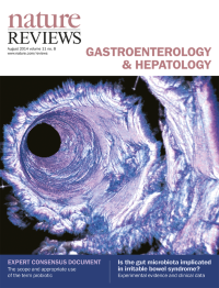
No. 8 August 2014
Cover image supplied by M. J. Gora, V. J. Madden and G. J. Tearney, Wellman Center for Photomedicine at Massachusetts General Hospital and Harvard Medical School, USA. A 3D image of the oesophagus created by rendering data obtained from an unsedated human subject using a swallowable tethered capsule endomicroscopy device. The capsule employs optical coherence tomography: optics within the capsule spin a focused beam around its circumference, acquiring cross-sectional images as it traverses the organ via peristalsis. A flexible tether containing an optical fibre is attached to the capsule and can be used to control its position and to remove it from the mouth so that it can be disinfected and reused.
-
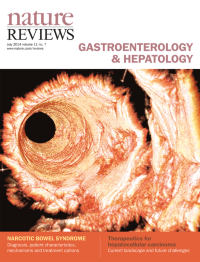
No. 7 July 2014
Cover image supplied by M. J. Gora, V. J. Madden and G. J. Tearney, Wellman Center for Photomedicine at Massachusetts General Hospital and Harvard Medical School, USA. A 3D image of the oesophagus created by rendering data obtained from an unsedated human subject using a swallowable tethered capsule endomicroscopy device. The capsule employs optical coherence tomography: optics within the capsule spin a focused beam around its circumference, acquiring cross-sectional images as it traverses the organ via peristalsis. A flexible tether containing an optical fibre is attached to the capsule and can be used to control its position and to remove it from the mouth so that it can be disinfected and reused.
-
No. 6 June 2014
Cover image supplied by M. J. Gora, V. J. Madden and G. J. Tearney, Wellman Center for Photomedicine at Massachusetts General Hospital and Harvard Medical School, USA. A 3D image of the oesophagus created by rendering data obtained from an unsedated human subject using a swallowable tethered capsule endomicroscopy device. The capsule employs optical coherence tomography: optics within the capsule spin a focused beam around its circumference, acquiring cross-sectional images as it traverses the organ via peristalsis. A flexible tether containing an optical fibre is attached to the capsule and can be used to control its position and to remove it from the mouth so that it can be disinfected and reused.
-
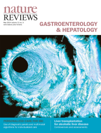
No. 5 May 2014
Cover image supplied by M. J. Gora, V. J. Madden and G. J. Tearney, Wellman Center for Photomedicine at Massachusetts General Hospital and Harvard Medical School, USA. A 3D image of the oesophagus created by rendering data obtained from an unsedated human subject using a swallowable tethered capsule endomicroscopy device. The capsule employs optical coherence tomography: optics within the capsule spin a focused beam around its circumference, acquiring cross-sectional images as it traverses the organ via peristalsis. A flexible tether containing an optical fibre is attached to the capsule and can be used to control its position and to remove it from the mouth so that it can be disinfected and reused.
-
No. 4 April 2014
Cover image supplied by M. J. Gora, V. J. Madden and G. J. Tearney, Wellman Center for Photomedicine at Massachusetts General Hospital and Harvard Medical School, USA. A 3D image of the oesophagus created by rendering data obtained from an unsedated human subject using a swallowable tethered capsule endomicroscopy device. The capsule employs optical coherence tomography: optics within the capsule spin a focused beam around its circumference, acquiring cross-sectional images as it traverses the organ via peristalsis. A flexible tether containing an optical fibre is attached to the capsule and can be used to control its position and to remove it from the mouth so that it can be disinfected and reused.
-
No. 3 March 2014
Cover image supplied by M. J. Gora, V. J. Madden and G. J. Tearney, Wellman Center for Photomedicine at Massachusetts General Hospital and Harvard Medical School, USA. A 3D image of the oesophagus created by rendering data obtained from an unsedated human subject using a swallowable tethered capsule endomicroscopy device. The capsule employs optical coherence tomography: optics within the capsule spin a focused beam around its circumference, acquiring cross-sectional images as it traverses the organ via peristalsis. A flexible tether containing an optical fibre is attached to the capsule and can be used to control its position and to remove it from the mouth so that it can be disinfected and reused.
-
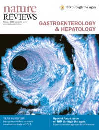
No. 2 February 2014
Cover image supplied by M. J. Gora, V. J. Madden and G. J. Tearney, Wellman Center for Photomedicine at Massachusetts General Hospital and Harvard Medical School, USA. A 3D image of the oesophagus created by rendering data obtained from an unsedated human subject using a swallowable tethered capsule endomicroscopy device. The capsule employs optical coherence tomography: optics within the capsule spin a focused beam around its circumference, acquiring cross-sectional images as it traverses the organ via peristalsis. A flexible tether containing an optical fibre is attached to the capsule and can be used to control its position and to remove it from the mouth so that it can be disinfected and reused.
Focus
-
No. 1 January 2014
Cover image supplied by M. J. Gora, V. J. Madden and G. J. Tearney, Wellman Center for Photomedicine at Massachusetts General Hospital and Harvard Medical School, USA. A 3D image of the oesophagus created by rendering data obtained from an unsedated human subject using a swallowable tethered capsule endomicroscopy device. The capsule employs optical coherence tomography: optics within the capsule spin a focused beam around its circumference, acquiring cross-sectional images as it traverses the organ via peristalsis. A flexible tether containing an optical fibre is attached to the capsule and can be used to control its position and to remove it from the mouth so that it can be disinfected and reused.