
Volume 3 Issue 11, November 2008
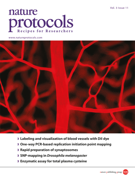
Retinal vasculature visualized by direct DiI labeling. A flat-mounted retina was viewed by fluorescence microscopy. The microphotograph shows a retinal artery with its branches. The nuclei of endothelial cells are identifiable as oval spots. Image is from the protocol by Li et al.
Protocol
-
Advertisement
