Thank you for visiting nature.com. You are using a browser version with limited support for CSS. To obtain
the best experience, we recommend you use a more up to date browser (or turn off compatibility mode in
Internet Explorer). In the meantime, to ensure continued support, we are displaying the site without styles
and JavaScript.
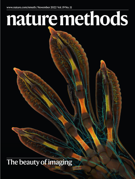
The winning image of the Nikon Small World 2022 Photomicrography Competition, an embryonic foot of a Madagascar giant day gecko (Phelsuma grandis). The image was captured using whole-mount fluorescence staining, tissue clearing, high-resolution confocal microscopy and image stitching.
Harvester ants live in desert grasslands and eat seeds. Colonies manage water stress by regulating foraging using olfactory interactions between outgoing and returning foragers. A long-term study in New Mexico shows how this collective behavior is evolving in drought conditions.
The EQIPD framework for rigor in animal experiments aims to unify current recommendations based on evidence behind their rationale and was prospectively tested for feasibility in multicenter animal experiments.
Researchers use electric fields to transfer RNA from a tissue sample onto a surface for subsequent fluorescence in situ hybridization-based profiling of transcriptomes at the single-cell level.
The generation of a whole larval zebrafish brain electron microscopy volume in tandem with automated tools lays the groundwork for producing the first vertebrate brain connectome.
An approach for integrating the wealth of heterogeneous brain data — from gene expression and neurotransmitter receptor density to structure and function — allows neuroscientists to easily place their data within the broader neuroscientific context.
RNA comprises a substantial fraction of eukaryotic chromatin, but techniques to identify and map RNAs are cumbersome. We adapted existing tagmentation-based profiling techniques to enable chromatin-associated RNAs to be profiled in a simple workflow, enhancing the capability to identify regulatory RNAs.
Light-Seq combines high resolution imaging with next generation sequencing of selected cell populations in fixed biological samples. Specifically, microscopically analyzed cells can be subjected to RNA expression profiling while keeping the sample intact for further assays, enabling cellular phenotypes and states to be assessed in the context of the original tissue.
A combination of light-sheet fluorescence microscopy (LSFM) with structured illumination doubles resolving power over LSFM alone. We show a practical implementation using a single objective for illumination and fluorescence detection and demonstrate its use for rapid volumetric imaging.
This Resource presents a serial block-face EM dataset of the whole larval zebrafish brain, including automated segmentation of neurons, detection of synapses and reconstruction of circuitry for visual motion processing.
SyConn2 is a machine learning-based framework for inferring and analyzing the connectomes contained in a volume electron microscopy dataset of brain tissue, for example from the zebra finch.
Iterative Synthetically Phosphorylated Isomers (iSPI) is a proteome-scale library of human-derived phosphoserine-containing phosphopeptides with precisely known positions of phosphorylation. This multi-purpose resource is available for optimization, standardization, and benchmarking of key steps in phosphoproteomics workflows.
This paper presents an iterative procedure where AlphaFold models are automatically rebuilt on the basis of experimental density maps and the rebuilt models are used as templates in new AlphaFold predictions.
Light-Seq uses light-directed DNA barcoding in fixed cells and tissues for multiplexed spatial indexing and subsequent next generation sequencing. This approach blends spatial and omics information to enable analysis of rare cell types in complex tissues.
ClampFISH 2.0 enables highly specific multiplexed signal amplification in RNA FISH. The approach was used to detect 10 RNA species that ranged in abundance in more than 1 million cells and is also applicable to tissue sections.
STELLAR (spatial cell learning) is a geometric deep learning model that works with spatially resolved single-cell datasets to both assign cell types in unannotated datasets based on a reference dataset and discover new cell types.
The longstanding goal of combining the optical sectioning of light-sheet illumination and the resolving power of multidirectional structured illumination microscopy is realized using an oblique plane microscope for improved fast 3D subcellular imaging.
Richardson–Lucy Network (RLN) combines the traditional Richardson–Lucy iteration with deep learning for improved deconvolution. RLN is more generalizable, offers fewer artifacts and requires less computing time than alternative approaches.
Omnipose is a deep neural network algorithm for image segmentation that improves upon existing approaches by solving the challenging problem of accurately segmenting morphologically diverse cells from images acquired with any modality.
cAMPFIREs are genetically encoded cAMP sensors that are suitable for in vivo imaging of cAMP signaling, as demonstrated in Drosophila larvae and behaving mice.