Thank you for visiting nature.com. You are using a browser version with limited support for CSS. To obtain
the best experience, we recommend you use a more up to date browser (or turn off compatibility mode in
Internet Explorer). In the meantime, to ensure continued support, we are displaying the site without styles
and JavaScript.
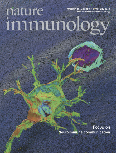
The nervous and immune systems communicate and reciprocally influence their functional responses. This month's joint focus presents review articles that examine how the nervous system and immune cells interact during development, homeostasis and in pathogenic disease states. http://www.nature.com/focus/neuroimmune_communication/index.html Artwork by Lewis Long.
We present a special set of Review articles on neuroimmune communication that highlight how the immune system and nervous system are anatomically connected, mechanistically communicate and reciprocally influence the other's function.
Enteric neurons and intestinal immune cells co-develop in response to common cues and communicate with each other to maintain organ function and host defense.
Engelhardt and colleagues review barriers separating blood from CSF and CNS parenchyma, how pathways draining solutes from CNS to lymph nodes exclude trafficking of antigen-presenting cells and how intravital microscopy has influenced debate on immune privilege of the CNS.
Various neurotrophic pathogens are capable of infecting CNS tissues. Klein and colleagues review how immune responses and inflammation in the CNS affect brain function and mental status.
The identification of VGLL3 as a transcription (co-)factor that underlies the sex bias of the human immune system further underscores the relevance of research into this area.
Regulatory T cells develop in the thymus as a distinct lineage of T cells instructed by the lineage-specifying transcription factor Foxp3. Epigenetic imprinting by the genome organizer Satb1 precedes this cell-fate 'decision' during thymocyte development.
Increased expression of the ubiquitin ligase TRAF6 in hematopoietic stem cells promotes activity of the GTP-binding protein Cdc42 and consequent diminished function of hematopoietic stem cells by ubiquitination of the RNA-binding protein hnRNP-A1, which leads to an inability to properly process pre-mRNA encoding Cdc42-inhibitory GTPase-activating proteins.
Various autoimmune diseases have sex-linked biases. Gudjonsson and colleagues find that the transcription factor VGLL3 is associated with a female-biased molecular signature linked to susceptibility to autoimmune disease.
Abramson and colleagues show that the coordinated action of several transcriptional regulators, including Irf4, Irf8, Tbx21, Tcf7 and Ctcfl, acts on medullary-thymic-epithelial-cell-specific accessible regions in the locus encoding the transcriptional regulator Aire to control its expression.
Thymic regulatory T (Treg) precursors undergo a distinct developmental pathway. Sakaguchi and colleagues show the chromatin organizer Satb1 is required for establishing the super-enhancer chromatin landscape of Treg cell–specific signature genes before Foxp3 expression.
Natural killer T cells in the thymus are CD1d-restricted cells that are selected at the CD4+CD8+ double-positive stage and require a variety of transcription factors for their development. Orkin, Winau and colleagues show that the histone demethylase UTX serves an essential role in the transcriptional control of the thymic maturation of these cells through multiple epigenetic mechanisms.
The phosphatase calcineurin targets NFAT transcription factors in T cells. Ashwell and colleagues show that calcineurin is recruited to the TCR signaling complex, where it reverses the inhibitory phosphorylation of the kinase Lck.
Cornall and colleagues show that Themis2 interacts with the phospholipase PLC-γ2 and lowers the threshold for B cell activation by low but not high avidity antigens.
Sensors of RNA viruses trigger prion-like aggregation of the adaptor MAVS, which leads to antiviral responses. Gao and colleagues show that the E3 ligase TRIM31 positively regulates this process by K63-linked polyubiquitination of MAVS.
Gringhuis, Geijtenbeek and colleagues show that the RNA helicase DDX3 binds abortive HIV-1 RNA and induces type I interferon in dendritic cells, a process that is inhibited by the HIV-1-induced activation of kinase PLK1.
Starczynowski and colleagues show that overexpression of TRAF6 in HSCs induces ubiquitination of the RNA-binding protein hnRNPA1 and alternative splicing of Arhgap1, which accounts for the hematopoietic defects in myelodysplastic syndromes.