Volume 10
-
No. 12 December 2009
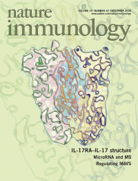
The interleukin 17 (IL-17) family includes five receptors and six cytokines, and both receptors and cytokines seem to form dimers. By solving the crystal structure of a complex between the receptor IL-17RA and an IL-17F homodimer, Garcia and colleagues (p 1245) provide insight into the molecular basis of IL-17 receptor heterodimerization. The original image shows a model of an IL-17F homodimer in blue, an IL-17RA molecule in yellow and an IL-17 receptor other than IL-17RA in pink. Original image by Lauren Ely and K. Christopher Garcia. Artwork by Lewis Long.
-
No. 11 November 2009
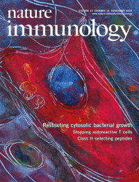
Bacteria that escape intracellular vacuoles and enter the cytoplasm of mammalian cells frequently become coated with polyubiquitin proteins. Randow and colleagues show that NDP52 binds these ubiquitin moieties and is required for the autophagy and restriction of cytoplasmic bacteria (p 1215 and News and Views by Ivanov and Roy, p 1137). The original confocal image shows colocalization (yellow) of samonella (green) and NDP52 (red) in HeLa cells. DAPI nuclear staining is blue. Original image by Felix Randow. Artwork by Lewis Long.
-
No. 10 October 2009
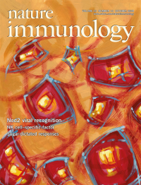
The pattern-recognition receptor Nod2 facilitates activation of transcription factor NF-κB in response to bacterial peptidoglycan. Bose and colleagues find that Nod2, together with the adaptor protein MAVS, is also required for virus-induced interferon-β production (p 1073 and News and Views by Murray, p 1053). The original confocal image shows colocalization (yellow) of Nod2 (red) and MAVS (green) in respiratory syncytial virus-infected primary normal human bronchial epithelial cells. Original image by Te Hung Chang and Victoria Frohlich. Artwork by Lewis Long.
-
No. 9 September 2009
Infection with human immunodeficiency virus type 1 (HIV-1) affects communication between immune cells. Cerutti and colleagues show that HIV-1-infected macrophages deliver the HIV-1 negative factor Nef through conduits to distal B cells, inhibiting their function (p 1008 and News and Views by Rudnicka & Schwartz, p 933). The original image by Dominika Rudnicka, Nathalie Sol-Foulon & Olivier Schwartz shows HIV-1-infected T cells immunostained for Nef (green), actin (red) and uninfected target T cells (blue). Artwork by Lewis Long.
-
No. 8 August 2009
Self-reactive thymocytes are purged by negative selection in the medulla of the thymus. Robey and colleagues demonstrate that autoreactive thymocytes migrate more slowly and in a more confined region than do polyclonal thymocytes (p 823; see also News and Views by Klein, p 809). The original image by Marie Le Borgne and Ena Ladi shows maximal projections of adjacent three-dimensional data sets spanning a cut thymus, with dendritic cells in yellow and blood vessels in red. Artwork by Lewis Long.
-
No. 7 July 2009
Basophils coexpress interleukin 4 and major histocompatibility complex class II molecules. Groups led by Artis, Nakanishi and Medzhitov (pp 697, 706 and 713; see also News and Views by Wynn, p 679) all report basophils as antigen-presenting cells that initiate T helper type 2 responses. Original collage shows a basophil isolated from a Schistosoma mansoni egg-infected 4get mouse with staining of major histocompatibility complex class II (red) and nuclei (blue) and endogenous expression of interleukin 4enhanced green fluorescent protein (green); lower right, merged image. Original images by Lingli Zhang and Jacqueline Perrigoue. Artwork by Lewis Long.
-
No. 6 June 2009
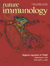
The MyD88-independent TLR4 pathway is mediated by the adaptor TRAM. O'Neill and colleagues (p 579) show that a splice variant of TRAM, called TAG, negatively regulates this pathway. The original image shows an HEK293 cell expressing fluorescence-tagged TLR4 (blue), TRAM (green) and the early endosome marker EEA1 (red). Cyan, yellow and purple represent the colocalization of TLR4 with TRAM, EEA1 with TRAM, and TLR4 with EEA1, respectively. Original image by Sarah. L. Doyle. Artwork by Lewis Long.
-
No. 5 May 2009
Experimental autoimmune encephalomyelitis requires the entry of disease-inducing T cells into the brain. Reboldi and colleagues (p 514; see also News and Views by Steinman, p 453) find that TH-17 cells initiate this disease by entering the brain through the choroid plexus. The original image shows human brain tissue in which choroid plexus epithelial cells are stained with antibody to CCL20 (fuchsia) and astrocytes are 'decorated' by antibody to glial fibrillary acidic protein (brown). Original image by Andrew Elston (LifeSpan BioSciences). Artwork by Lewis Long.
-
No. 4 April 2009
Long thought to travel in straight paths from membrane receptors to the nucleus, many immune receptor signaling cascades actually follow more convoluted pathways, some of which become entangled with signals emanating from heterologous receptors. In this issue, a series of specially commissioned articles discuss the molecular basis for and biological consequences of this dynamic signaling interaction. Additional insights on immune signaling cross-talk are available online (http://www.nature.com/ni/focus/crosstalk/index.html). Artwork by Lewis Long.
-
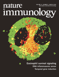
No. 3 March 2009
Prosurvival cytokines prevent activation and mitochondrial localization of the apoptosis-inducing factor Bax. Malter and colleagues (p 257) find that the peptidyl-prolyl isomerase Pin1 is integral to this process. The original confocal image shows activated Bax (green) partially colocalized (yellow) with mitochondria (red). Original image by Z.-J. Shen. Artwork by Lewis Long.
-
No. 2 February 2009
Inflammation triggered by oxygen deprivation or hypoxia can complicate clinical procedures such as organ transplantation. Eltzschig and colleagues (p 195) find that inflammation induced by hypoxia is restrained by netrin-1, which blocks neutrophil transmigration. The original image shows the expression of netrin-1 (green) induced by hypoxia in mouse intestine (blue indicates DAPI nuclear staining). Original image by M. Faigle. Artwork by Lewis Long.
-
No. 1 January 2009
Nonsignaling chemokine receptors such as DAR C are thought to function as chemokine 'decoys'. Rot and colleagues (p 101) show that DAR C transports chemokines unidirectionally to the apical face of endothelial barriers. The original micrograph shows massive infiltration of inflammatory leukocytes to sites of cutaneous hypersensitivity in DAR C-transgenic mice. Original image by Monika Pruenster, Paula Bombosi and Antal Rot. Artwork by Lewis Long.