Thank you for visiting nature.com. You are using a browser version with limited support for CSS. To obtain
the best experience, we recommend you use a more up to date browser (or turn off compatibility mode in
Internet Explorer). In the meantime, to ensure continued support, we are displaying the site without styles
and JavaScript.
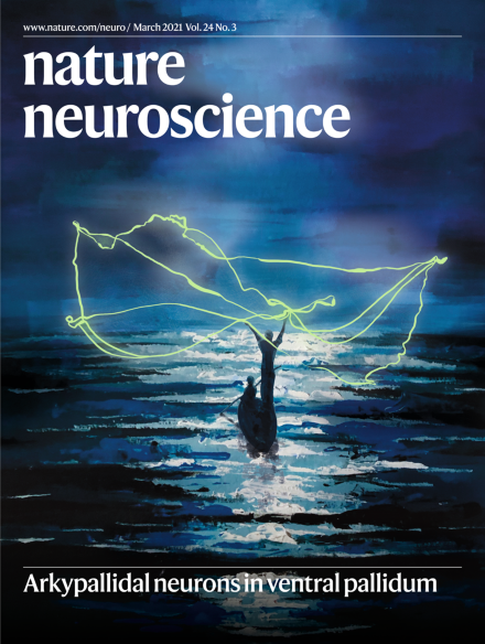
The cover art is an artistic rendition of arkypallidal (vArky) neurons, which originate in the ventral pallidum. Their name comes from the ancient Greek ἄρκυς [arkys] for “hunter's net” (Mallet et al., 2012), from their filigree-like processes that extend throughout the accumbens and exert broad nets of influence over neural activity.
Leslie Gail Ungerleider, a distinguished experimental psychologist and neuroscientist, previously Chief of the Laboratory of Brain and Cognition at the National Institute of Mental Health, died suddenly on 11 December 2020. Friends, family, colleagues, and trainees all the world over mourn her passing, but also celebrate her life and extraordinary achievements.
A new study discovered that ventral pallidal neurons projecting back to the nucleus accumbens promote consumption. The findings call into question the accepted direction of information flow through the ventral basal ganglia and open new avenues for studying how consumption is regulated in proportion to subjective value.
By probing the dependence on learning history or motivational significance of stimuli, Sharpe and colleagues reveal previously uncharacterized contributions of lateral hypothalamus GABA neurons to associative learning.
Gene-based therapies offer the promise of long-lasting clinical benefit for both genetic and sporadic neurodegenerative diseases. Sun and Roy highlight recent successes and caveats, offering a prospective glimpse into this rapidly emerging arena.
Good–bad binary classifications fail to describe reactive astrocytes in CNS disorders. Here, 81 researchers reach consensus on widespread misconceptions and provide definitions and recommendations for future research on reactive astrocytes.
Gava et al. explore the organization of neuronal co-activity in hippocampus from a graph theoretical perspective to report how new associative memories integrate into the network and restructure the neural patterns representing prior memories.
Gordon et al. use genome-wide unbiased approaches to show that human cerebral cortical organoids, when cultured for many months, start to resemble stages of postnatal brain development, with a timeline that parallels in vivo development.
Guttikonda et al. engineered a human pluripotent stem cell-derived tri-culture system containing microglia, astrocytes, and neurons. This system recapitulates cell-type-specific inflammatory signaling in an in vitro model of Alzheimer’s disease.
Synapse loss is prominent in the cortex in multiple sclerosis (MS). In a cortical MS model, Jafari et al. show that phagocytes remove synapses by engulfment, which is triggered by local calcium accumulations and prevented by blocking colony-stimulating factor 1 signaling.
Rhea at al. show that intravenously injected, radiolabeled SARS-CoV-2 spike 1 protein crosses the mouse blood–brain barrier, likely through the mechanism of adsorptive transcytosis and is also taken up by peripheral tissues.
Inhibition of nucleus accumbens neurons is crucial for reward consumption. Vachez, Tooley et al. characterize arkypallidal neurons in the ventral pallidum that inhibit accumbal neurons to sustain reward consumption in a value-dependent manner.
Sharpe et al. find that prior reward-learning experience can prime reward circuits to encode fear memories. This suggests prior experience can shape the way we learn, opening the neural boundaries for learning about particular types of information.
Spikes of deep-layer ID2+Nkx2.1+ cortical neurons are anticorrelated with spiking of all principal cells and interneurons, prominently during down states of sleep, and shape the sequential firing of neurons at down–up transitions.
Examination of neural activity reveals that performing a rapid sequence of actions depends not upon fusing those actions into a holistic unit, but upon the ability of motor cortex to swiftly prepare the next action while the present unfolds.
This study defined spatial gene expression in the human dorsolateral prefrontal cortex. It reveals layer-enriched expression of genes associated with schizophrenia and autism, highlighting the clinical relevance of spatially defined expression.
Boulting et al. profile activity-dependent gene expression and regulatory elements in human induced pluripotent stem cell-derived GABAergic neurons and uncover a possible role for calcium-responsive gene promoters of these neurons in autism risk.