Volume 24
-
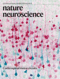
No. 12 December 2021
Corticospinal motor circuitsNelson et al. reveal how the widespread axon collaterals of corticospinal neurons are organized throughout the nervous system, and how projections to the striatum and spinal cord are coordinated through cell type-specific connectivity. The cover art depicts virally labeled corticospinal neurons in blue and corticostriatal neurons in red.
See Nelson et al.
-
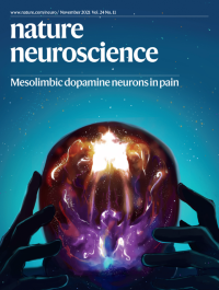
No. 11 November 2021
Mesolimbic dopamine neurons in painThe cover art depicts the agony of negative emotional states accompanying persistent pain. The bright yellow and purple spots represent the mesolimbic dopamine pathway, which becomes less active in pain, leading to negative emotional states such as decreased motivation and anhedonia. The presence of these negative states can often lead to feelings of isolation, represented as an individual in the vastness of space.
See Markovic et al.
-
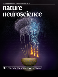
No. 10 October 2021
EEG marker for seizure onset zoneThe primary treatment for drug-resistant epilepsy is surgical removal of brain regions causing seizures, but outcomes are poor because there is no biomarker of epileptogenicity. Li and colleagues developed an EEG marker, neural fragility, that pinpoints brain regions likely to cause seizures. The cover art illustrates fragile (orange) epileptogenic neurons on the chalice triggering a seizure, represented by flames and smoke, resulting in a hyperactive EEG (light blue traces).
See A. Li et al.
-
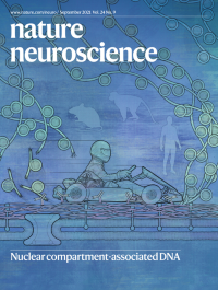
No. 9 September 2021
Nuclear compartment-associated DNAAhanger and colleagues map 3D genome architecture associated with two nuclear compartments—the lamina and the nuclear speckles—in the developing mammalian brain using Genome Organization with CUT and RUN Technology (GO-CaRT). The cover art illustrates protein A–micrococcal nuclease, depicted here as an actual go-cart, racing along the antibody-defined nuclear lamina and tearing up any chromatin in its path.
See S. H. Ahanger et al.
-
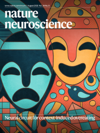
No. 8 August 2021
Neural circuit for context-induced overeatingFood-related cues in the environment can trigger overeating. Mohammad and colleagues reveal a neural circuit that promotes this non-homeostatic feeding driven by environmental context. The cover art illustrates that the same food items, depicted here as lollipops and candy bars, can stimulate or lead to loss of appetite depending on the context.
See Mohammad et al
-
No. 7 July 2021
Resolving uncertainty in hunger and thirstEiselt et al. report limitations on the ability of mice to assess their hunger and thirst states under conditions that model human decisions. Incorrect food-seeking in thirst may contribute to overeating. The cover illustrates the uncertainty of a thirsty mouse deciding between food and water.
See Eiselt et al.
-
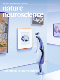
No. 6 June 2021
Multimodal neural recordingsThis cover features an electrode -- Neuro-FITM (flexible, insertable, and transparent microelectrode) – walking through a gallery of brain images (wide-field imaging) while listening to the audio guide, trying to understand the meaning of the artworks (brain activity). The transparent clothes of the electrode represent the high transparency of Neuro-FITM.
See Liu et al
-
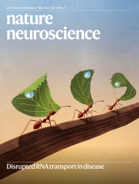
No. 5 May 2021
Disrupted RNA transport in diseaseRNAs often travel great distances before they are translated into proteins. Fernandopulle et al. review how neurons orchestrate long-range RNA transport and how disruptions in this process contribute to neurological disease. The cover depicts a series of ants carrying dew-laden leaves, echoing the way that RNA granules—which exist as liquid droplets—indirectly move along microtubule networks.
See Fernandopulle et al.
-
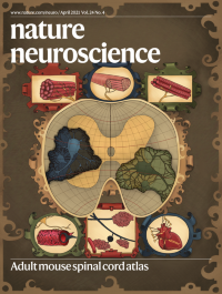
No. 4 April 2021
Adult mouse spinal cord atlasBlum and colleagues reveal the transcriptional and functional diversity of motor neuron subtypes within the spinal cord. The cover art illustrates these findings using a classical world atlas rendered as a spinal cord cross-section, where skeletal and autonomic motor neurons reside on two main continents but extend their reach to distal muscle fibers and effector cells throughout the body.
See Blum et al.
-
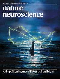
No. 3 March 2021
Arkypallidal neurons in ventral pallidumThe cover art is an artistic rendition of arkypallidal (vArky) neurons, which originate in the ventral pallidum. Their name comes from the ancient Greek ἄρκυς [arkys] for “hunter's net” (Mallet et al., 2012), from their filigree-like processes that extend throughout the accumbens and exert broad nets of influence over neural activity.
See Vachez et al
-
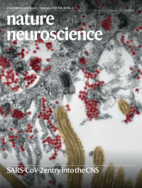
No. 2 February 2021
SARS-CoV-2 entry into the CNSMeinhardt, Radke and Dittmayer et al. identify intact coronavirus particles by electron microscopy within the olfactory mucosa of an individual with COVID-19. SARS-CoV-2 particles (colored in red) are visible within and above a ciliated cell (kinocilia in yellow) in the vicinity of olfactory receptor neurons (not depicted here).
See Meinhardt et al.
-
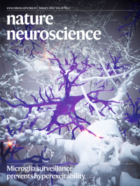
No. 1 January 2021
Microglia surveillance prevents hyperexcitabilityMicroglia constantly extend and retract processes to survey the brain parenchyma. Merlini, Rafalski and colleagues report the role of dynamic microglial brain surveillance in the prevention of brain network hyperexcitability. The cover art illustrates microglia processes contacting neuronal somata to reduce excessive neuronal activity and prevent neuronal hypersynchrony and seizures.
See Merlini et al.